- Health Hub
- /
- scans
A guide to pregnancy ultrasounds: types & what to expect
scans
5 minutes reading time
Let's face it: pregnancy is a lot. On top of all the planning, learning, nesting, and sickness, you also have to go to OB/GYN appointments, where doctors hit you with a lot of overwhelming information and advice. The whole process can feel impersonal, strange, and sometimes a bit scary. Knowing what to expect during your pregnancy can help you navigate it with more confidence. Here's a helpful breakdown of what you can expect when it comes to ultrasounds.
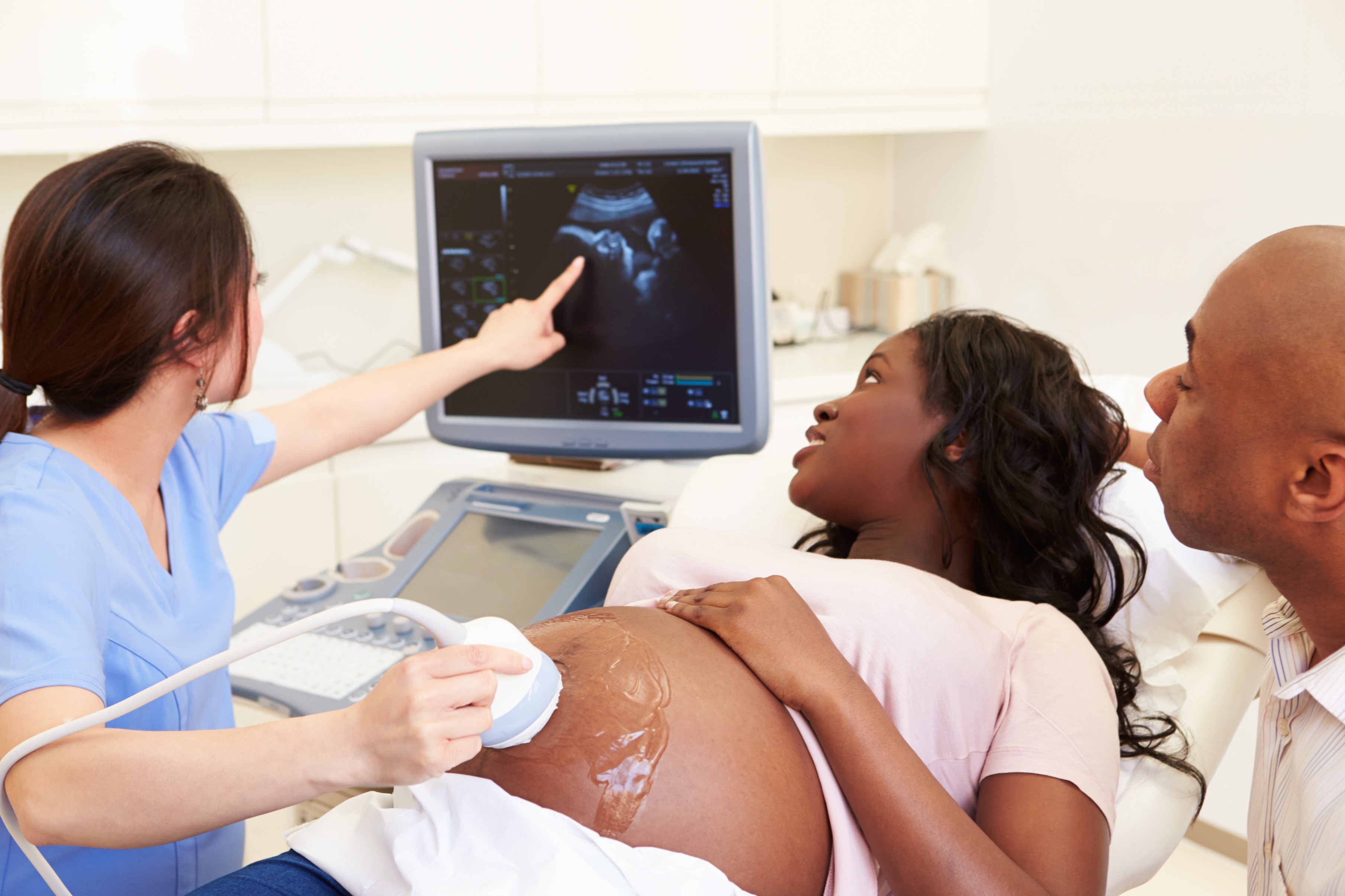
Ultrasounds are medical imaging tests that can be used to detect and confirm pregnancies. Transducers are the instruments used for ultrasound exams. In pregnancy, they work by sending out high-frequency sound waves that create images of your reproductive system.
Before we dive into the nuances of ultrasounds, you'll need a crash course on pregnancy math. You would think that when you're late for your period because you're pregnant, you're maybe a few days or weeks along, right? Wrong. According to the bizarre laws of pregnancy math, you are already 4 weeks pregnant at the time of your missed period. If you’re a week late, you're 5 weeks pregnant. If you're 4 weeks late, you're 8 weeks pregnant. Crazy, huh?
Your first ultrasound probably won’t be scheduled until you are 8 weeks pregnant (and remember, that’s 4 weeks after your missed period!), though pregnancy can be detected by ultrasound as soon as a few days after a fertilized egg implants in the uterus.1 However, doctors typically won’t schedule ultrasounds that early on because the chance of miscarriage before the 8-week mark is significant.
Some facilities call the first ultrasound a “pregnancy confirmation” or a “level one ultrasound”, while others simply call it a “viability scan”. (For as exciting and emotionally charged as pregnancy can be, the terminology people use at its various stages can be curiously cold and sterile.)
In early pregnancy, your first ultrasound will likely be a transvaginal one. During a transvaginal ultrasound, a trained technician will gently insert a transducer wand into your vagina to capture clear images of your uterus, cervix, ovaries, and fallopian tubes. You will feel pressure, but no pain. The images will confirm whether your pregnancy is intrauterine (a fetus has implanted in your uterus) or extrauterine (a fetus has implanted outside your uterus, also known as an ectopic pregnancy).
In most ectopic pregnancies, a fertilized egg implants in a fallopian tube; in others, it implants in an ovary, a cervix, or elsewhere in the abdominal cavity. A fertilized egg cannot survive outside the uterus, so an ectopic pregnancy will, sadly, end in miscarriage. Ultrasounds are critical in detecting ectopic pregnancies, which can burst fallopian tubes and cause life-threatening internal bleeding.2
When getting images of your uterus, the technician doing your ultrasound will record the fetus's heartbeat and measure its size. The physical measurements of the fetus tell the technician its gestational age, which helps them calculate approximately how far along your pregnancy is. This can be especially helpful in the event you have irregular periods or are unsure of when you conceived.
Your approximate due date will be calculated based on the gestational age of the fetus. (Note: if the fetus is fetuses in your case, if your pregnancy is considered high risk or has unusual circumstances of any kind, your ultrasounds, testing, monitoring, and other medical needs may vary from those described in this article.)
An early pregnancy ultrasound doesn’t always look how you might think. The white shape on the screen might look much more like a cashew, a gummy bear, or a tiny alien than a baby. Early in an intrauterine pregnancy, you will likely also see a white circle near the embryo. That's a yolk sac, and its job is to provide important nutrients to the fetus until the placenta takes over that role. (In case you didn’t know, you're going to grow a whole new organ around the 10- to 12-week mark. Like, from scratch.)
The other two standard ultrasounds happen at weeks 13 and 20 weeks. Both ultrasounds are done trans-abdominally, using the gel and handheld transducer you’ve probably seen on movies and TV. The 13-week ultrasound, sometimes referred to as the nuchal translucency scan, takes about 30 to 45 minutes, depending on the baby’s position and visibility.
The scan’s purpose is to confirm developmental milestones and mark potential abnormalities. For the first time, you will be able to see the baby’s brain, heart, organs, bones, and the width of its neck. You might even get to see it move around! This ultrasound also shows which uterine wall your placenta has attached to (front, back, top, or bottom).
If your technician doesn’t mention the placenta’s location, be sure to ask! Knowing if it’s in the front (anterior) or the back (posterior) of your uterus will give you an indication of how easily felt fetal kicks and movement will be once they start in a month or two. (The placenta acts like a thin cushion between you and your baby. If the placenta is in the back, you’ll feel movement more easily. If it’s in the front, the movement might be more difficult to detect.)
The 20-week ultrasound, more popularly referred to as the “anatomy scan”, is the one where technicians can make a birth-sex declaration, and generally takes about 45 to 60 minutes. Its purpose is to capture highly detailed images of the baby’s bones, major organs, and spinal cord, and spot certain birth defects. You will be able to see your baby’s fingers, toes, beating heart, and even its facial bones quite clearly during this ultrasound.
The technician will be checking your placental position, your amniotic fluid volume, and your cervical length as well. They may do a transvaginal ultrasound if they need a closer or more detailed view of your cervix. If everything looks and measures normal, the 20-week ultrasound may be the last ultrasound you have during your pregnancy. If abnormalities are suspected or detected in the 13- or 20-week ultrasound, your doctor will talk to you about it and order more tests to monitor the baby’s development.
While that can be a scary thing to hear, additional tests are often ordered for relatively minor reasons, like poor visibility due to the baby’s position, or pockets of lower-than-optimal amniotic fluid. Plus, more ultrasounds mean more chances to see your baby before it’s born. Helpful hint: be sure to go into both of these scans with a full bladder–it will push your uterus further away from your lower pelvis and make it easier for the technician to get clear images.
Growing a new human in your body is an incredible but mysterious experience. Ultrasounds are so valuable because they take pictures in great detail, allowing your doctor to provide you and your baby the best care possible from conception to delivery.
You can find an ultrasound scan center near you by using scan.com’s scan search tool.
Resources: