1. Health Hub
  2. /
  3. scans
04 May 2022

What's in the pipeline? Six promising medical imaging trends in 2022

scans 3 minutes reading time

It’s an exciting time in medicine. Over the last decade, industry-disruptors like gene editing, 3D printing, and artificial intelligence have entered the mainstream of medical treatment. Diagnostic technology is evolving at a fast clip, too.

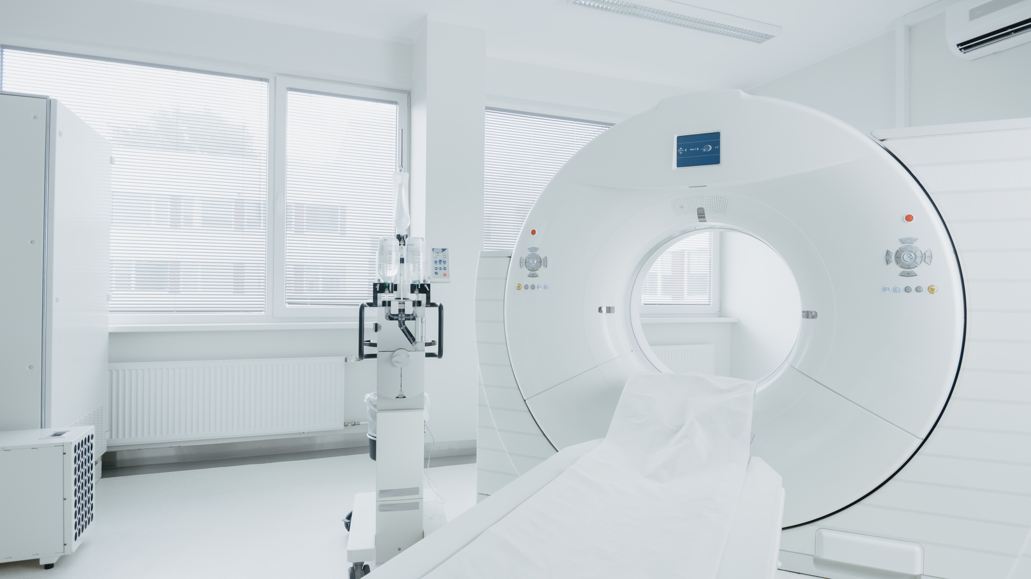
Medical imaging scans like X-rays, MRIs, CT scans, PET scans, mammograms, and ultrasounds make up most of the current industry standard for diagnosing or confirming disease, injury, or abnormality. However, the level of detail and precision in each of these scans is not always optimal, and new, innovative imaging technologies designed to improve patient diagnoses and outcomes are emerging all the time.

Six medical imaging trends to follow in 2022

Here are a few exciting developments in the world of medical imaging this year:

  1. Artificial intelligence and machine learning (AI and ML). In recent years, AI and ML have augmented radiological workflow using advanced detection methods. AI can quickly spot a major stroke and route the scan to the top of the radiologist’s imaging pile, allowing for quicker diagnosis and emergency treatment in a narrow therapeutic window.1 AI and ML also improve day-to-day procedural efficiency for radiologists who are poring over high volumes of scans each day. Soon, AI and ML may be able to use deep-learning algorithms to mine data for new disease markers. They will also likely play a major role in the shift of imaging and clinical data to the cloud in the near future, streamlining healthcare information in a way that will benefit healthcare providers and patients alike.
  2. Photon counting detector (PCD) technology. PCD is considered next-generation computed tomography (CT) technology, and works by sorting the energies from each X-ray once they’ve passed through a scan field.2 One X-ray with PCD can create multiple sets of CT data with higher precision visualization and finer segmentation than radiologists are used to seeing in standard CT scan images. A mobile PCD CT would be especially helpful in point-of-care emergency and surgical situations, where quick access to high-accuracy imaging can save a life.
  3. Prostate specific membrane antigen (PSMA) PET scans. Named one of the top 10 medical innovation watches in 2022 by Cleveland Clinic, PSMA PET scans are slated to become the new standard of care for diagnosing prostate cancer and detecting prostate cancer metastases.3 PMSA is a prostate cancer biomarker which is found in high levels on cancer surface cells. Prostate cancer is the most common form of cancer in men, particularly in the aging male population, accounting for 200,000 American cases annually. As it stands, conventional prostate imaging scans like MRIs and CTs have limits in terms of accuracy and clarity. This novel method of using radioactive tracers on PMSA proteins and visualizing the cells using CT and MRI could lead to improved care and outcomes for prostate cancer patients.
  4. Electromagnetic acoustic (EMA) imaging. EMA combines radio signals and sound waves to produce detailed, real-time visualizations of fluid accumulation, calcification, unhealthy tissue, and capillaries in a way that ultrasounds alone currently cannot do (ultrasounds only use sound waves).4 EMA stands out from current medical imaging scans in an important way: it can be used on the spot, during a doctor’s appointment (immediate point of care), to accurately image and diagnose a medical problem. This bypasses the process of having to get a separate referral to an imaging center and the anxious experience of waiting for your results.
  5. 3D mammography. Digital breast tomosynthesis (DBT) systems have been steadily replacing traditional 2D mammography units for years now, and are approaching a 50% presence in breast imaging centers in 2022. Traditional 2D preventive mammograms take X-ray images from two angles, whereas 3D mammography systems work more like computed tomography (CT) scans, taking a high number of slice-by-slice images that together create a composite picture. They’re expensive, but they allow radiologists to differentiate dense breast tissue from malignant tissue more easily. They also tend to reduce the number of biopsies and callbacks women will need after their preventive or diagnostic screenings.
  6. 3D lung X-rays. Sweden is in the process of testing a first-in-kind, high resolution, 3D X-ray image of lungs breathing in real time. Already, scientists have been able to see the thinnest and most microscopic structures in mice lungs as the mice inhale and exhale. This is a big deal because most imaging scans can only produce clear images when the patient is completely still. Here, the natural movement of breathing does not sacrifice any image clarity. This imaging technique utilizes a very small dose of radiation, which will make it low risk for humans once approved.

Resources:

  1. med-technews.com: Latest Medtech News - Med-Tech Innovation

  2. itnonline.com: Imaging Technology News

  3. radiologybusiness.com: Cleveland Clinic names PSMA PET a top 10 medical innovation to watch in 2022

  4. jacr.org: Journal of the American College of Radiology

Find an imaging center near you

Search for local MRI imaging centers near you and book online in minutes.