How it works
Click on highlighted words to see explanations!
How it works
Click on highlighted words to see explanations.

MRI Lumbar Spine Without Contrast
October 31, 2026

Click on highlighted words to see explanations!
MRI LUMBAR SPINE WITHOUT CONTRAST
CLINICAL INDICATION:
Low back pain radiating to the left leg for 6 weeks. Evaluate for disc herniation and nerve root compression.
TECHNIQUE:
Multiplanar, multisequence MRI of the lumbar spine was performed on a 1.5T magnet without intravenous contrast, including sagittal T1, sagittal T2, sagittal STIR, and axial T1 and T2 sequences.
COMPARISON:
None available.
FINDINGS:
For the purposes of this report, five lumbar-type vertebral bodies are assumed.
Alignment: Normal lumbar lordosis. No spondylolisthesis.
Vertebral Bodies: Vertebral body heights are maintained. No fracture.
Mild type 2 Modic endplate changes at L5-S1. No suspicious marrow lesion.
Conus Medullaris: Terminates normally at the L1 level. Normal signal.
Disc Levels:
T12-L1: No disc herniation, spinal canal stenosis, or neural foraminal narrowing.
L1-L2: No disc herniation, spinal canal stenosis, or neural foraminal narrowing.
L2-L3: Mild disc desiccation. No disc herniation. No spinal canal stenosis or neural foraminal narrowing.
L3-L4: Mild disc bulge and mild bilateral facet arthropathy. No significant spinal canal stenosis. Mild bilateral neural foraminal narrowing.
L4-L5: Disc desiccation with mild loss of disc height. Left paracentral disc extrusion extending 6 mm posteriorly and 4 mm caudally, contacting and posteriorly displacing the traversing left L5 nerve root in the lateral recess. Moderate bilateral facet arthropathy and ligamentum flavum thickening. Mild-to-moderate spinal canal stenosis. Mild left neural foraminal narrowing. No right neural foraminal narrowing.
L5-S1: Disc desiccation and moderate loss of disc height. Broad-based disc bulge without focal herniation. Mild bilateral facet arthropathy.
No spinal canal stenosis. Mild bilateral neural foraminal narrowing without impingement of the exiting L5 nerve roots.
Paraspinal Soft Tissues: Unremarkable.
IMPRESSION:
1. L4-L5 left paracentral disc extrusion contacting and displacing the traversing left L5 nerve root, correlating with the patient's left leg symptoms.
2. Mild-to-moderate spinal canal stenosis at L4-L5.
3. Multilevel degenerative disc disease and facet arthropathy, most pronounced at L4-L5 and L5-S1.
4. Mild bilateral neural foraminal narrowing at L3-L4 and L5-S1.
Electronically signed by: Dr. Demo Radiologist, MD
Date/Time: 10/02/2026 09:47
Patient-friendly report powered by

MRI
MRI stands for “magnetic resonance imaging.” This is a type of radiology scan that uses magnets to create images of the body by measuring tiny changes in water molecules within the body’s tissues.
MRI
MRI
MRI का मतलब है “मैग्नेटिक रेजोनेंस इमेजिंग।” यह एक प्रकार का रेडियोलॉजी स्कैन है जो शरीर के ऊतकों के भीतर पानी के अणुओं में छोटे परिवर्तनों को मापकर शरीर की छवियां बनाने के लिए चुंबक का उपयोग करता है।
MRI
IRM
Abreviatura de Imagen de Resonancia Magnética. La resonancia magnética es un tipo de exploración radiológica que utiliza imanes para crear imágenes del cuerpo midiendo pequeños cambios en las moléculas de agua dentro de los tejidos del cuerpo.
MRI
MRI
MRI是”磁共振成像”的缩写。这是一种利用磁铁通过测量人体组织内水分子的微小变化来生成人体图像的放射学扫描。
MRI
IRM
IRM signifie « imagerie par résonance magnétique » Il s’agit d’un type d’examen radiologique qui utilise des aimants pour créer des images du corps en mesurant de minuscules changements dans les molécules d’eau à l’intérieur des tissus du corps.
LUMBAR SPINE
The lower back. The lumbar spine starts just below the rib cage and goes down to about the level of the hip bones. There are usually five lumbar vertebrae (spine bones).
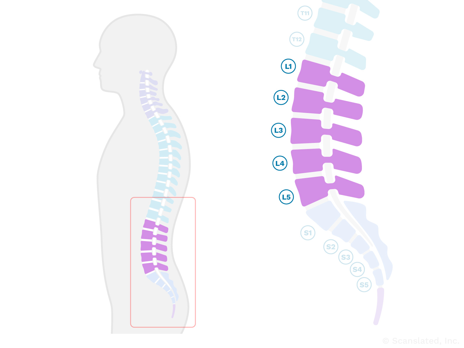
LUMBAR SPINE
काठ की रीढ़
पीठ के निचले हिस्सा। काठ का रीढ़ पसलियों के पिंजरे के ठीक नीचे से शुरू होता है और कूल्हे की हड्डियों के स्तर तक जाता है। सामान्यतः पांच कटि कशेरुकाएं (रीढ़ की हड्डियां) होती हैं।
LUMBAR SPINE
columna lumbar
la parte baja de la espalda. La parte inferior de la columna vertebral (justo por debajo de la caja torácica hasta aproximadamente el nivel de los huesos de la cadera) se denomina columna lumbar. Normalmente hay 5 huesos en la columna lumbar (vértebras).
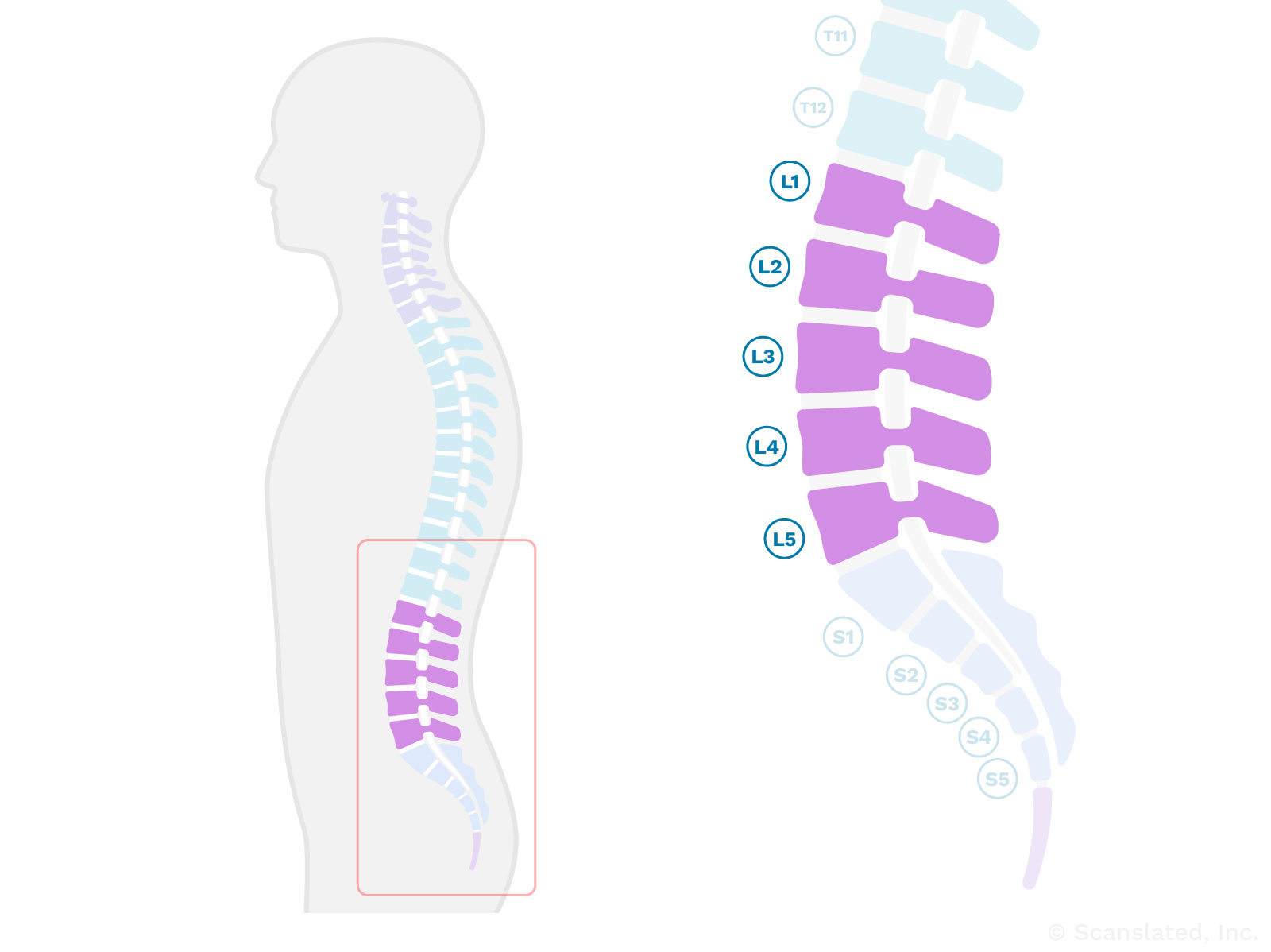
LUMBAR SPINE
腰椎
下背部。腰椎从胸廓的正下方开始, 一直延伸到大约髋骨的水平。通常有五块腰椎骨(椎骨)。
LUMBAR SPINE
colonne lombaire
Bas du dos. La colonne lombaire commence juste en dessous de la cage thoracique et descend jusqu’au niveau des os de la hanche. Il y a généralement cinq vertèbres lombaires (os de la colonne vertébrale).
WITHOUT CONTRAST
A scan that was performed without injecting any contrast dye (X-ray or MRI dye). Many types of scans do not need contrast dye, so this is normal.
Doing a scan without contrast dye can help the radiologist see certain things. It can also serve as a good comparison to a scan taken after contrast dye is injected, because it makes it easier to determine if something that looks bright on the scan is due to dye, or was already there before the dye was given.
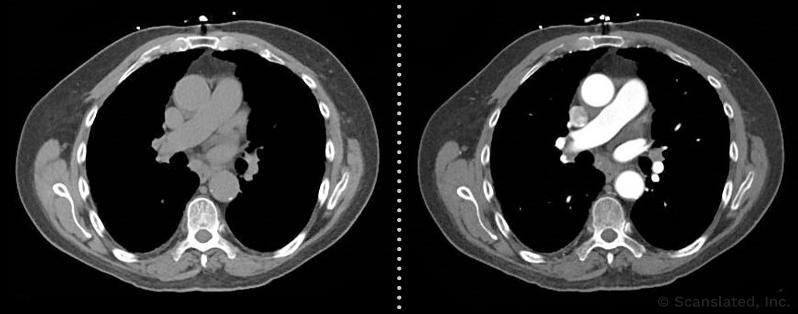
The image on the left was taken without contrast, and the one on the right was taken with contrast. These examples are NOT images from your exam.
WITHOUT CONTRAST
बिना कंट्रास्ट
एक स्कैन जो किसी भी कंट्रास्ट डाई (एक्स-रे या एमआरआई डाई) को इंजेक्ट किए बिना किया गया था। कई प्रकार के स्कैन में कॉन्ट्रास्ट डाई की आवश्यकता नहीं होती, इसलिए यह सामान्य है।
बिना कॉन्ट्रास्ट डाई के स्कैन करने से रेडियोलॉजिस्ट को कुछ चीजें देखने में मदद मिल सकती है। यह कंट्रास्ट डाई इंजेक्ट करने के बाद लिए गए स्कैन की तुलना में एक अच्छी तुलना के रूप में भी काम कर सकता है, क्योंकि इससे यह निर्धारित करना आसान हो जाता है कि स्कैन पर जो कुछ चमकीला दिखता है वह डाई के कारण है, या डाई लगने से पहले ही मौजूद था। दिया गया।
The image on the left was taken without contrast, and the one on the right was taken with contrast. These examples are NOT images from your exam.
WITHOUT CONTRAST
sin contraste
imágenes obtenidas sin ningún tipo de tinte
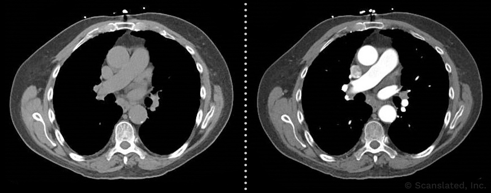
The image on the left was taken without contrast, and the one on the right was taken with contrast. These examples are NOT images from your exam.
WITHOUT CONTRAST
无造影剂
在不注射任何造影剂(X 光或核磁共振成像染料)的情况下进行的扫描。许多类型的扫描都不需要造影剂, 所以这是正常的。
在不使用造影剂的情况下进行扫描可以帮助放射科医生看清某些东西。它还可以与注射造影剂后进行的扫描进行比较, 因为这样可以更容易地确定扫描中看起来明亮的东西是造影剂造成的, 还是在注射造影剂之前就已经存在。
The image on the left was taken without contrast, and the one on the right was taken with contrast. These examples are NOT images from your exam.
WITHOUT CONTRAST
sans contraste
Imagerie réalisée sans injection de produit de contraste (colorant pour radiographie ou IRM). De nombreuses imageries ne nécessitent pas de produits de contraste, donc c’est normal.
Faire une imagerie sans produit de contraste peut aider le radiologue à voir certaines choses. Elle peut aussi servir de comparaison avec une image prise après injection d’un produit de contraste, car cela permet de déterminer plus aisément si quelque chose qui à l’air plus clair sur l’image l’est à cause du produit ou si cette chose était déjà claire avant que le produit soit injecté.
The image on the left was taken without contrast, and the one on the right was taken with contrast. These examples are NOT images from your exam.
CLINICAL INDICATION
The reason for the scan (the problem that led to your doctor ordering the scan)
leg
In common usage, “leg” refers to the entire lower limb (from the thigh down to the foot).
But, in medical terminology, “leg” refers to only the section from the knee to the foot.
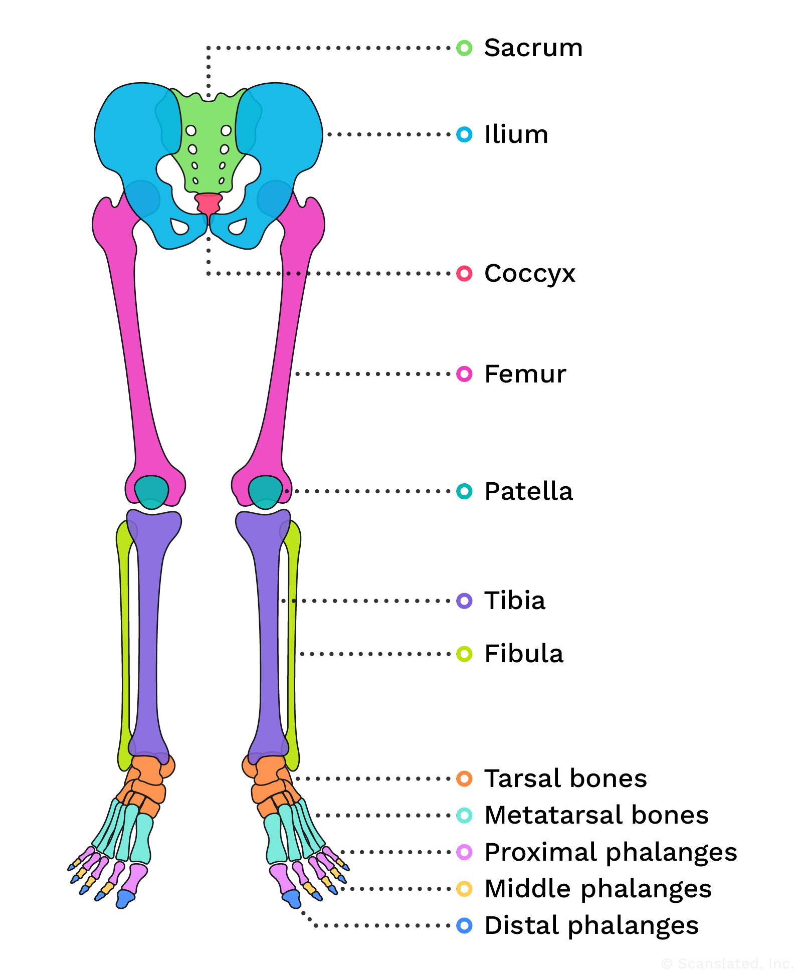
leg
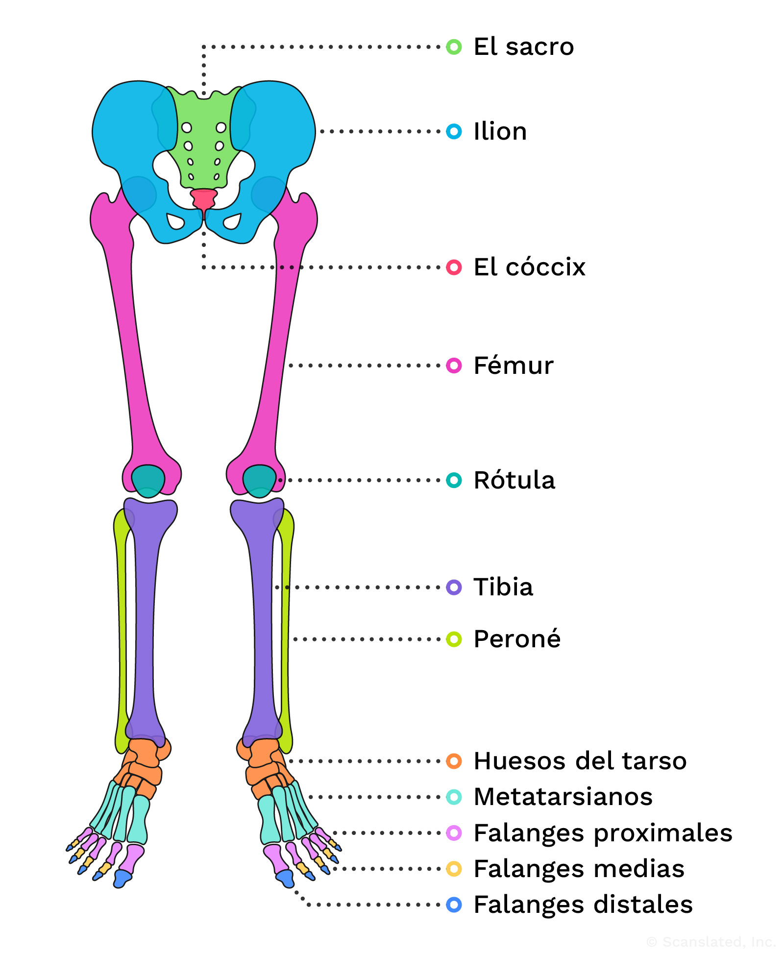
pierna
la parte del miembro inferior que va de la rodilla al pie
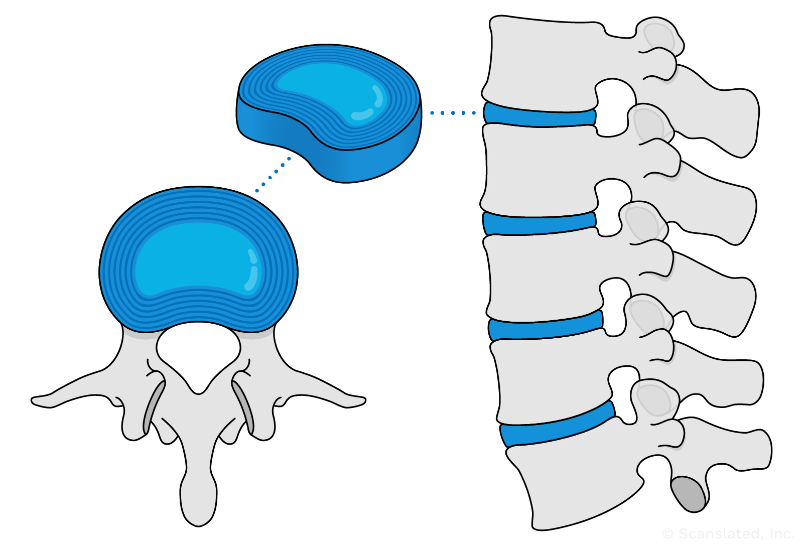
disc
Spinal discs (or “intervertebral discs”) are the soft, shock-absorbing cushions between spine bones (vertebrae) that allow the spine to flex.
disc
डिस्क
स्पाइनल डिस्क रीढ़ की हड्डियों के बीच नरम, आघात-अवशोषित कुशन होते हैं जो रीढ़ को लचीला होने देते हैं।
disc
disco
Los discos vertebrales son el material blando que amortigua los impactos entre los huesos de la columna vertebral.
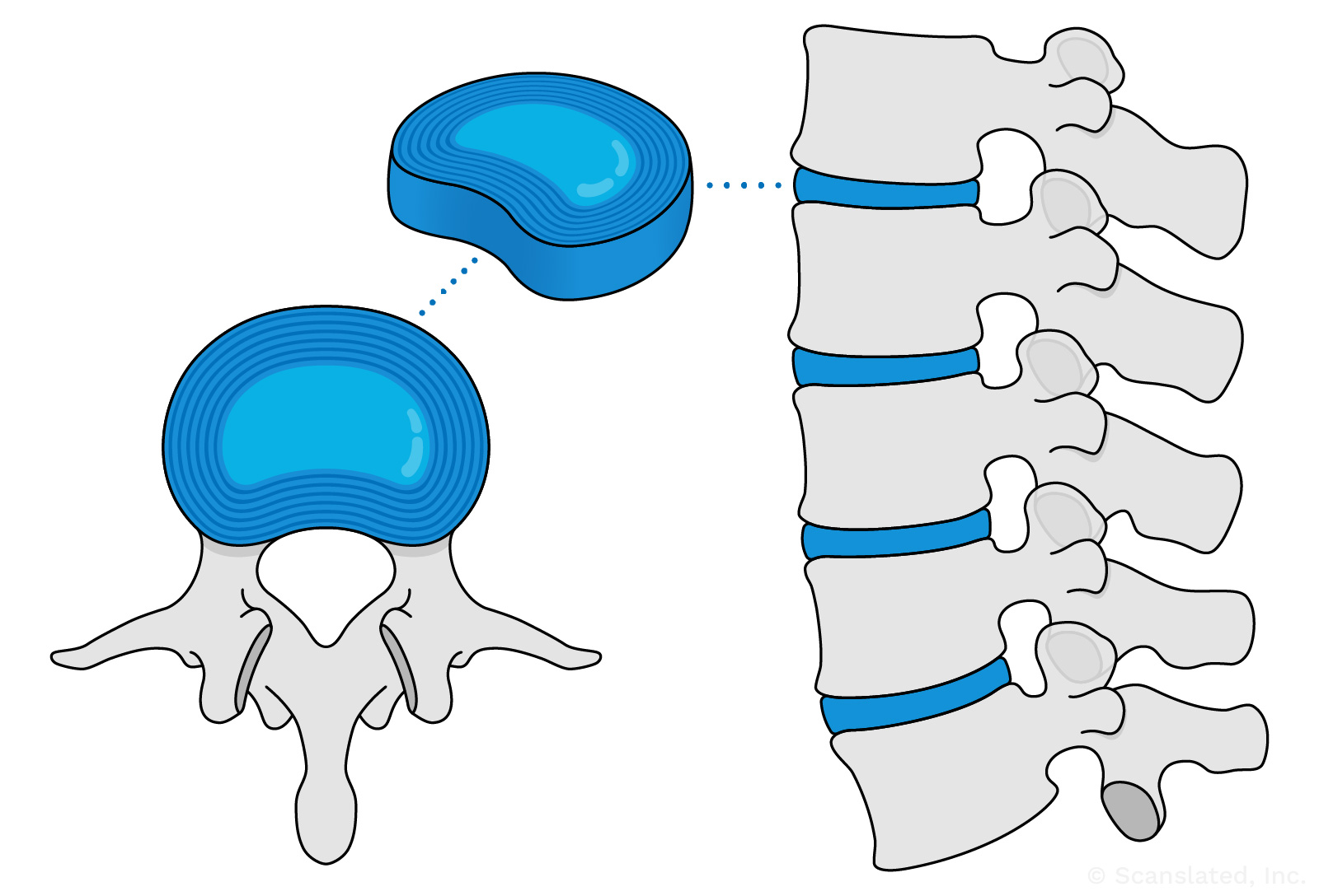
disc
椎间盘
椎间盘是椎骨之间的减震软垫, 使脊柱能够弯曲。
disc
disque
Les disques vertébraux (ou « disques intervértébraux ») sont des coussins souples qui absorbent les chocs entre les os de la colonne vertébrale (les vertèbres) et qui permettent à cette dernière de fléchir.
nerve root
The beginning part of a nerve, just as it’s leaving the spine to go out to the body
nerve root
तंत्रिका मूल
तंत्रिका का आरंभिक भाग, जैसे ही वह रीढ़ की हड्डी को छोड़कर शरीर में जाती है
nerve root
raíz nerviosa
La parte inicial de un nervio, justo cuando está saliendo de la columna vertebral para salir al cuerpo.
nerve root
神经根
神经的起始段, 即刚从脊柱伸出的一段, 其后延伸至身体各部位
nerve root
racine nerveuse
Partie initiale d’un nerf, au moment où il quitte la colonne vertébrale pour se diriger vers le corps.
TECHNIQUE
Refers to how an imaging test was performed
Multiplanar
Images created from several different perspectives (such as a side view, top view, front view, etc.)
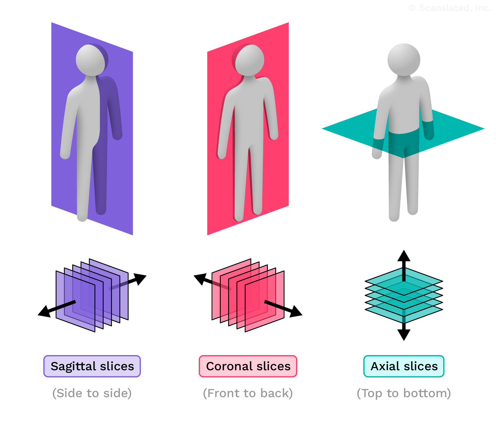
multisequence
Multiple different types of MRI images created by changing the settings on the machine.
When you have an MRI scan, the scanner will take multiple sets of images using different settings. Each set is called a “sequence,” and shows the body in a different way.
Looking at multiple sequences of the same body part gives the radiologist more information to work with when looking at the scan images.
multisequence
बहुअनुक्रम
मशीन की सेटिंग्स में परिवर्तन करके विभिन्न प्रकार की एमआरआई छवियां बनाई जा सकती हैं।
जब आप MRI स्कैन कराते हैं, तो स्कैनर अलग-अलग सेटिंग्स का उपयोग करके छवियों के कई सेट लेगा। प्रत्येक सेट को “अनुक्रम” कहा जाता है, और यह शरीर को अलग-अलग तरीके से दिखाता है।
एक ही शरीर के अंग के एकाधिक अनुक्रमों से रेडियोलॉजिस्ट को स्कैन छवियों को देखते समय काम करने के लिए अधिक जानकारी मिलती है।
multisequence
multisecuencial
Varios tipos de resonancia magnética. Se crean diferentes tipos de imágenes (llamadas secuencias) cambiando los ajustes de la máquina.
multisequence
多序列的
通过改变机器上的设置, 生成多种不同类型的MRI图像。
进行MRI扫描时, 扫描仪会使用不同的设置拍摄多组图像。每一组被称为一个”序列”, 以不同的方式展示身体。
通过查看同一身体部位的多个序列, 放射科医生可以在查看扫描图像时获得更多信息以进行分析。
multisequence
multiséquence
Différents types d’images IRM créées en modifiant les paramètres de l’appareil.
Lorsque vous passez une IRM, le scanner prend plusieurs séries d’images en utilisant différents paramètres. Chaque série est appelée « séquence » et montre le corps d’une manière différente.
L’examen de plusieurs séquences de la même partie du corps permet au radiologue de disposer de plus d’informations lorsqu’il examine les images du scanner.
MRI
MRI stands for “magnetic resonance imaging.” This is a type of radiology scan that uses magnets to create images of the body by measuring tiny changes in water molecules within the body’s tissues.
MRI
MRI
MRI का मतलब है “मैग्नेटिक रेजोनेंस इमेजिंग।” यह एक प्रकार का रेडियोलॉजी स्कैन है जो शरीर के ऊतकों के भीतर पानी के अणुओं में छोटे परिवर्तनों को मापकर शरीर की छवियां बनाने के लिए चुंबक का उपयोग करता है।
MRI
IRM
Abreviatura de Imagen de Resonancia Magnética. La resonancia magnética es un tipo de exploración radiológica que utiliza imanes para crear imágenes del cuerpo midiendo pequeños cambios en las moléculas de agua dentro de los tejidos del cuerpo.
MRI
MRI
MRI是”磁共振成像”的缩写。这是一种利用磁铁通过测量人体组织内水分子的微小变化来生成人体图像的放射学扫描。
MRI
IRM
IRM signifie « imagerie par résonance magnétique » Il s’agit d’un type d’examen radiologique qui utilise des aimants pour créer des images du corps en mesurant de minuscules changements dans les molécules d’eau à l’intérieur des tissus du corps.
lumbar spine
The lower back. The lumbar spine starts just below the rib cage and goes down to about the level of the hip bones. There are usually five lumbar vertebrae (spine bones).
lumbar spine
काठ की रीढ़
पीठ के निचले हिस्सा। काठ का रीढ़ पसलियों के पिंजरे के ठीक नीचे से शुरू होता है और कूल्हे की हड्डियों के स्तर तक जाता है। सामान्यतः पांच कटि कशेरुकाएं (रीढ़ की हड्डियां) होती हैं।
lumbar spine
columna lumbar
la parte baja de la espalda. La parte inferior de la columna vertebral (justo por debajo de la caja torácica hasta aproximadamente el nivel de los huesos de la cadera) se denomina columna lumbar. Normalmente hay 5 huesos en la columna lumbar (vértebras).
lumbar spine
腰椎
下背部。腰椎从胸廓的正下方开始, 一直延伸到大约髋骨的水平。通常有五块腰椎骨(椎骨)。
lumbar spine
colonne lombaire
Bas du dos. La colonne lombaire commence juste en dessous de la cage thoracique et descend jusqu’au niveau des os de la hanche. Il y a généralement cinq vertèbres lombaires (os de la colonne vertébrale).
without intravenous contrast
A scan that was performed without injecting any contrast dye (X-ray or MRI dye). Many types of scans do not need contrast dye, so this is normal.
Doing a scan without contrast dye can help the radiologist see certain things. It can also serve as a good comparison to a scan taken after contrast dye is injected, because it makes it easier to determine if something that looks bright on the scan is due to dye, or was already there before the dye was given.
The image on the left was taken without contrast, and the one on the right was taken with contrast. These examples are NOT images from your exam.
without intravenous contrast
बिना अंतःशिरा कंट्रास्ट के
एक स्कैन जो किसी भी कंट्रास्ट डाई (एक्स-रे या एमआरआई डाई) को इंजेक्ट किए बिना किया गया था। कई प्रकार के स्कैन में कॉन्ट्रास्ट डाई की आवश्यकता नहीं होती, इसलिए यह सामान्य है।
बिना कॉन्ट्रास्ट डाई के स्कैन करने से रेडियोलॉजिस्ट को कुछ चीजें देखने में मदद मिल सकती है। यह कंट्रास्ट डाई इंजेक्ट करने के बाद लिए गए स्कैन की तुलना में एक अच्छी तुलना के रूप में भी काम कर सकता है, क्योंकि इससे यह निर्धारित करना आसान हो जाता है कि स्कैन पर जो कुछ चमकीला दिखता है वह डाई के कारण है, या डाई लगने से पहले ही मौजूद था। दिया गया।
The image on the left was taken without contrast, and the one on the right was taken with contrast. These examples are NOT images from your exam.
without intravenous contrast
sin contraste intravenoso
imágenes obtenidas sin ningún tipo de tinte
The image on the left was taken without contrast, and the one on the right was taken with contrast. These examples are NOT images from your exam.
without intravenous contrast
无静脉造影剂增强
在不注射任何造影剂(X 光或核磁共振成像染料)的情况下进行的扫描。许多类型的扫描都不需要造影剂, 所以这是正常的。
在不使用造影剂的情况下进行扫描可以帮助放射科医生看清某些东西。它还可以与注射造影剂后进行的扫描进行比较, 因为这样可以更容易地确定扫描中看起来明亮的东西是造影剂造成的, 还是在注射造影剂之前就已经存在。
The image on the left was taken without contrast, and the one on the right was taken with contrast. These examples are NOT images from your exam.
without intravenous contrast
sans produit de contraste par intraveineuse
Imagerie réalisée sans injection de produit de contraste (colorant pour radiographie ou IRM). De nombreuses imageries ne nécessitent pas de produits de contraste, donc c’est normal.
Faire une imagerie sans produit de contraste peut aider le radiologue à voir certaines choses. Elle peut aussi servir de comparaison avec une image prise après injection d’un produit de contraste, car cela permet de déterminer plus aisément si quelque chose qui à l’air plus clair sur l’image l’est à cause du produit ou si cette chose était déjà claire avant que le produit soit injecté.
The image on the left was taken without contrast, and the one on the right was taken with contrast. These examples are NOT images from your exam.
sagittal
In CT or MRI scans, “sagittal” refers to images that are oriented as a side view, showing slices through the body from side to side.
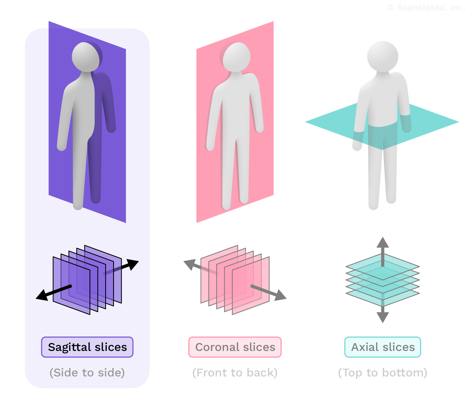
sagittal
धनु
सीटी या एमआरआई स्कैन में, “सेगिटल” से तात्पर्य उन छवियों से है जो पार्श्व दृश्य के रूप में होती हैं, तथा शरीर के एक ओर से दूसरी ओर के हिस्सों को दिखाती हैं।
sagittal
sagital
imágenes que se orientan como una proyección lateral a través del cuerpo
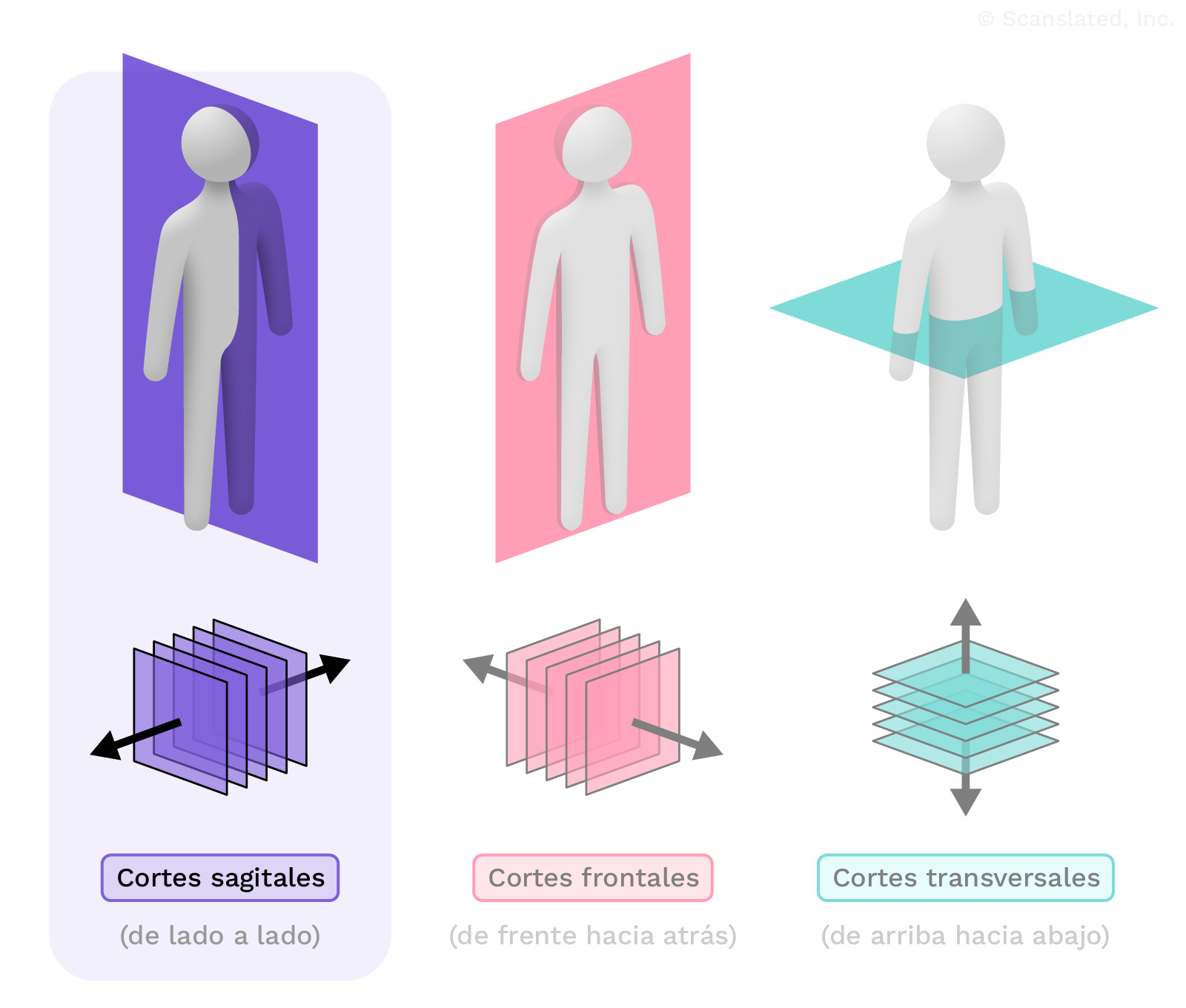
sagittal
矢状位
在 CT 或核磁共振成像扫描中, “矢状位”指的是从侧面视图方向拍摄影像, 显示从人体一侧到另一侧的切层。
sagittal
sagittal(e)
En tomodensitométrie ou en IRM, le terme « sagittal » désigne les images orientées depuis un côté, montrant des coupes du corps d’un côté à l’autre.
sagittal
In CT or MRI scans, “sagittal” refers to images that are oriented as a side view, showing slices through the body from side to side.
sagittal
धनु
सीटी या एमआरआई स्कैन में, “सेगिटल” से तात्पर्य उन छवियों से है जो पार्श्व दृश्य के रूप में होती हैं, तथा शरीर के एक ओर से दूसरी ओर के हिस्सों को दिखाती हैं।
sagittal
sagital
imágenes que se orientan como una proyección lateral a través del cuerpo
sagittal
矢状位
在 CT 或核磁共振成像扫描中, “矢状位”指的是从侧面视图方向拍摄影像, 显示从人体一侧到另一侧的切层。
sagittal
sagittal(e)
En tomodensitométrie ou en IRM, le terme « sagittal » désigne les images orientées depuis un côté, montrant des coupes du corps d’un côté à l’autre.
sagittal
In CT or MRI scans, “sagittal” refers to images that are oriented as a side view, showing slices through the body from side to side.
sagittal
धनु
सीटी या एमआरआई स्कैन में, “सेगिटल” से तात्पर्य उन छवियों से है जो पार्श्व दृश्य के रूप में होती हैं, तथा शरीर के एक ओर से दूसरी ओर के हिस्सों को दिखाती हैं।
sagittal
sagital
imágenes que se orientan como una proyección lateral a través del cuerpo
sagittal
矢状位
在 CT 或核磁共振成像扫描中, “矢状位”指的是从侧面视图方向拍摄影像, 显示从人体一侧到另一侧的切层。
sagittal
sagittal(e)
En tomodensitométrie ou en IRM, le terme « sagittal » désigne les images orientées depuis un côté, montrant des coupes du corps d’un côté à l’autre.
STIR
Stands for “short tau inversion recovery,” which is a common type of MRI image that makes it easier to see fluid in the tissues (like with swelling or injury).
On these images, fluid appears very bright, and everything else appears dark.
STIR
हलचल
इसका अर्थ है “शॉर्ट टाऊ इनवर्सन रिकवरी”, जो कि एमआरआई छवि का एक सामान्य प्रकार है, जो ऊतकों में तरल पदार्थ (जैसे सूजन या चोट के साथ) को खोजना आसान बनाता है।
इन चित्रों में तरल पदार्थ बहुत चमकीला दिखाई देता है, तथा शेष सब कुछ अंधकारमय दिखाई देता है।
STIR
STIR
Significa “Recuperación de la Inversión Tau Corta”. Es un tipo de resonancia magnética habitual que facilita la visualización del líquido en los tejidos.
STIR
STIR
指的是”短时反转恢复序列”, 这是一种常见的MRI图像类型, 可以更容易地看到组织中的液体(例如用于肿胀或受伤)。
在这种图像中, 液体显得非常明亮, 其他部分则显得很暗。
STIR
remuer
Il s’agit d’un type courant d’image IRM qui facilite la visualisation des fluides dans les tissus (comme dans le cas d’un gonflement ou d’une blessure).
Sur ces images, le fluide apparaît très brillant et tout le reste est sombre.
sequences
Different types of MRI images created by changing the settings on the machine.
When you have an MRI scan, the scanner will take multiple sets of images using different settings. Each set is called a “sequence,” and shows the body in a different way.
Looking at multiple sequences of the same body part gives the radiologist more information to work with when looking at the scan images.
sequences
अनुक्रम
मशीन की सेटिंग्स में परिवर्तन करके विभिन्न प्रकार की एमआरआई छवियां बनाई गईं।
जब आप एमआरआई स्कैन कराएंगे, तो स्कैनर विभिन्न सेटिंग्स का उपयोग करके छवियों के कई सेट लेगा। प्रत्येक सेट को “अनुक्रम” कहा जाता है, और यह शरीर को अलग-अलग तरीके से दिखाता है।
एक ही शरीर के अंग के कई अनुक्रमों को देखने से रेडियोलॉजिस्ट को स्कैन छवियों को देखते समय काम करने के लिए अधिक जानकारी मिलती है।
sequences
secuencias
Tipos de IRM creados por la modificación de los ajustes en la máquina. Cuando se realiza una resonancia magnética, la máquina tomará múltiples conjuntos de imágenes utilizando diferentes configuraciones. Estos ajustes diferentes se denominan secuencias y cada una de ellas muestra el cuerpo de una manera diferente. La observación de varias secuencias (o tipos de imágenes) de la misma parte del cuerpo aumenta la cantidad de información que se puede obtener al observar las imágenes de la exploración.
sequences
序列
通过改变仪器上的设置生成不同类型的核磁共振成像影像。
进行核磁共振成像扫描时, 扫描仪会使用不同的设置拍摄多组影像。每一组影像被称为一个”序列”, 以不同的方式展示身体。
放射科医生在查看扫描影像时, 通过查看同一身体部位的多个序列可以获得更多信息。
sequences
séquences
Différents types d’images IRM créés en modifiant les paramètres de la machine.
Quand vous passez une IRM, le scanner prend plusieurs jeux d’images avec différents paramètres. Chaque jeu est appelé « séquence » et montre le corps de différente manière.
L’examen de plusieurs séquences de la même partie du corps permet au radiologue de disposer de plus d’informations lorsqu’il examine les images du scanner.
COMPARISON:
This section of the report lists any previous imaging tests (X-rays, scans, etc.) that are available.
Radiologists may compare old images with the new ones to see if anything has changed.
COMPARISON:
तुलना:
रिपोर्ट के इस भाग में उपलब्ध पूर्व इमेजिंग परीक्षणों (एक्स-रे, स्कैन, आदि) की सूची दी गई है।
रेडियोलॉजिस्ट पुरानी छवियों की तुलना नई छवियों से करके देख सकते हैं कि क्या कुछ परिवर्तन हुआ है।
COMPARISON:
comparación
En esta sección del informe se enumeran las disponibles pruebas de diagnóstico por la imagen anteriores (radiografías, exploraciones). Es posible que los médicos comparen las imágenes antiguas con las nuevas para determinar si algo ha cambiado.
COMPARISON:
对比:
报告的这一部分列出了可用的先前任何影像检查(X 光、扫描等)。
放射科医生可能会将旧影像与新影像进行比较, 以确定是否有任何变化。
COMPARISON:
comparaison :
Cette section du compte rendu énumère tous les examens d’imagerie antérieurs (radiographies, scanners, etc.) disponibles.
Les radiologues peuvent comparer les anciennes images avec les nouvelles pour voir si quelque chose a changé.
None available
There were no previous X-rays or scans available to compare with.
None available
कोई उपलब्ध नहीं
तुलना के लिए कोई पूर्व एक्स-रे या स्कैन उपलब्ध नहीं था।
None available
No hay ninguno disponible
Esto quiere decir que no habían radiografías o exploraciones antiguas con las que comparar.
None available
不可用
没有先前的 X 光片或扫描结果可供比较。
None available
aucun(e) disponible
Il n’y a pas de radiographie antérieure disponible à des fins de comparaison.
FINDINGS
“Findings” refers to the observations that were made when looking at the images.
vertebral bodies
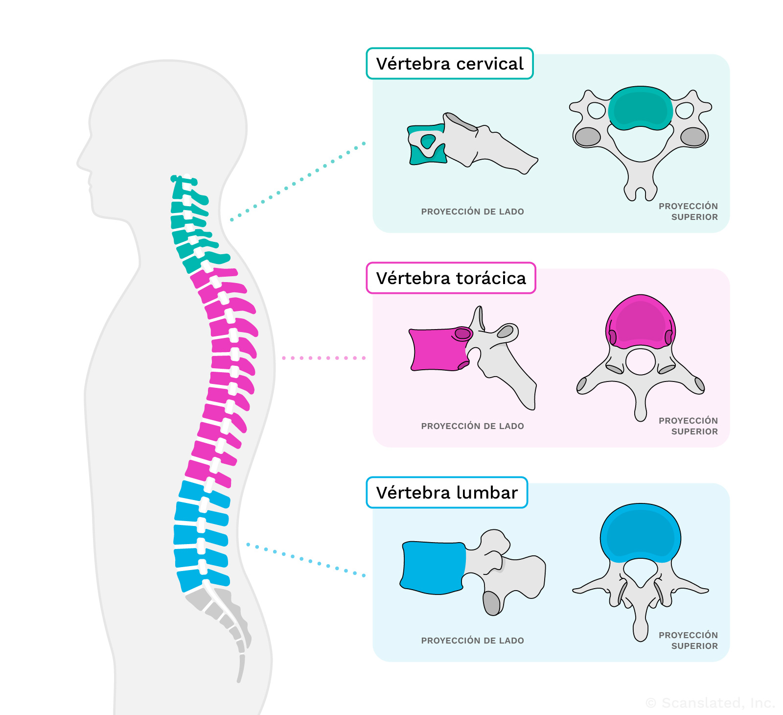
The cube-shaped portions of the spine bones (“vertebrae”), which stack to form the spinal column. Between vertebrae there are discs, which are soft structures that cushion the bones. The discs help to support the weight of the body and to absorb impacts.
The spine is separated into four segments:
- Cervical (the neck)
- Thoracic (through the area of the chest)
- Lumbar (the lower back)
- Sacral (the crescent-shaped bones in between your hip bones that you sit on)
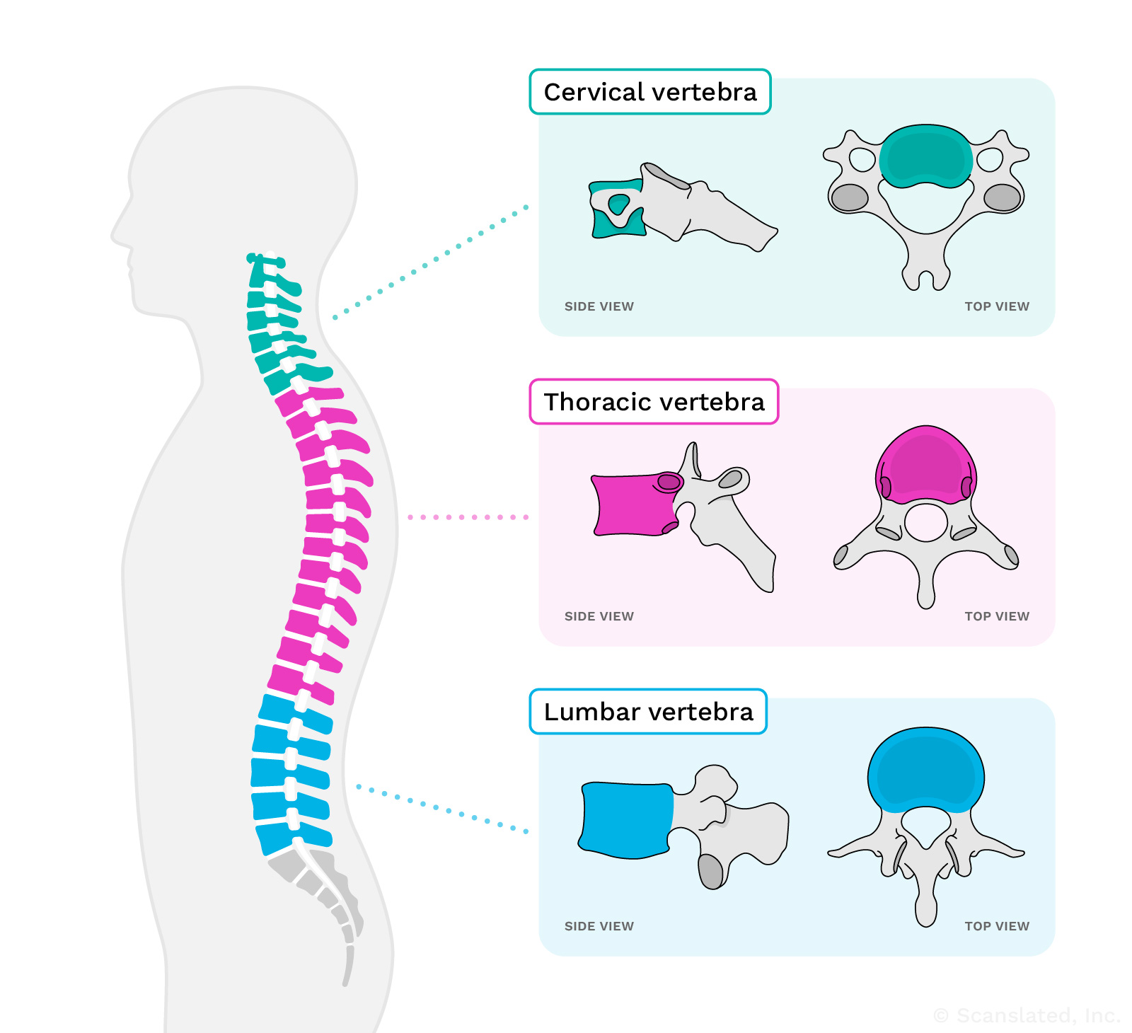
vertebral bodies
कशेरुका निकाय
रीढ़ की हड्डियों (“कशेरुक”) के घनाकार भाग, जो एकत्रित होकर मेरुदण्ड बनाते हैं। कशेरुकाओं के बीच में नरम संरचनाएं होती हैं जो हड्डियों को सहारा देती हैं जिन्हें डिस्क कहा जाता है। डिस्क शरीर के वजन को सहारा देने और आघातों को अवशोषित करने में मदद करती हैं।
रीढ़ की हड्डी को चार खंडों में विभाजित किया गया है:
– सरवाइकल (गर्दन) - वक्षीय (छाती के क्षेत्र से होकर) - लम्बर (पीठ के निचले हिस्से) - सैक्रल (आपके कूल्हे की हड्डियों के बीच की अर्धचंद्राकार हड्डियाँ जिन पर आप बैठते हैं)
vertebral bodies
Cuerpos vertebrales
Los cuerpos vertebrales son los huesos en forma de cubo/cuadrado de la espalda (hay 33 de ellos desde el cuello hasta la parte baja de la espalda). Los cuerpos vertebrales están apilados uno encima del otro para formar la columna vertebral. Estos huesos tienen proyecciones que se extienden hacia atrás y forman un anillo óseo, por donde pasan la médula espinal y los nervios.
vertebral bodies
椎体
脊椎骨(”椎骨”)的立方体部分, 它们堆叠在一起形成脊柱。椎骨之间有椎间盘, 它们是软性结构, 用来缓冲骨骼。椎间盘有助于支撑身体的重量和吸收冲击力。
脊柱分为四个部分:
- 颈椎(颈部)
- 胸椎(通过胸部区域)
- 腰椎(下背部)
- 骶椎(位于髋骨之间的弯月形骨骼, 您就坐在上面)
vertebral bodies
corps vertébraux
Les parties en forme de cube des os de la colonne vertébrale (« vertèbres »), qui s’empilent pour former la colonne vertébrale. Entre les vertèbres se trouvent des disques, structures souples qui amortissent les os. Les disques aident à supporter le poids du corps et à absorber les chocs.
La colonne vertébrale est séparée en quatre segments :
- Cervical (le cou)
- Thoracique (dans la région de la poitrine)
- Lombaire (le bas du dos)
- Sacral (les os en forme de croissant situés entre les os de la hanche et sur lesquels vous vous asseyez)
Alignment
The way two bones are lined up with one another at a joint or in the spine.
Normally, bones are lined up perfectly. When a joint is dislocated, the bones are offset and no longer line up together.
Alignment
संरेखन
जिस प्रकार दो हड्डियां एक जोड़ पर या रीढ़ की हड्डी में एक दूसरे के साथ पंक्तिबद्ध होती हैं।
आम तौर पर, हड्डियाँ पूरी तरह से पंक्ति में रहतीं हैं। जब कोई जोड़ खिसक जाता है, तो हड्डियां एक दूसरे से अलग हो जाती हैं और एक पंक्ति में नहीं रहतीं।
Alignment
alineación
La forma en que dos huesos se alinean entre sí en una articulación.Por lo general, los huesos están perfectamente alineados.Cuando una articulación se disloca, los huesos se desplazan y dejan de estar alineados.
Alignment
对齐
两块骨头在关节处或脊柱中相互排列的方式。
正常情况下, 骨骼是完全排列整齐的。当关节脱位时, 骨骼会偏移, 不再排在一起。
Alignment
alignement
La façon dont deux os sont alignés l’un par rapport à l’autre au niveau d’une articulation ou de la colonne vertébrale.
Normalement, les os sont parfaitement alignés. Lorsqu’une articulation est disloquée, les os sont décalés et ne sont plus alignés.
Normal lumbar lordosis
The lower back has its normal curvature.
The cervical (neck) and lumbar (lower back) sections of the spine normally have a slight curvature called a “lordosis.”
Normal lumbar lordosis
सामान्य लम्बर लॉर्डोसिस
पीठ के निचले हिस्से में सामान्य वक्रता होती है।
रीढ़ की हड्डी के ग्रीवा (गर्दन) और काठ (पीठ के निचले हिस्से) भागों में सामान्यतः हल्का सा वक्रता होता है, जिसे “लॉर्डोसिस” कहा जाता है।
Normal lumbar lordosis
lordosis lumbar normal
La ligera curvatura normal hacia atrás que se observa en la parte baja de la espalda. Es normal tener esta curva gradual hacia atrás en la columna lumbar.
Normal lumbar lordosis
正常的腰椎前凸
下背部有一个通常正常的弧度。
颈椎(颈部)和腰椎(腰部)通常有一个轻微的弯曲, 称为”前凸”。
Normal lumbar lordosis
lordose lombaire normale
Le bas du dos présente la courbure habituelle et normale.
Les sections cervicale (cou) et lombaire (bas du dos) de la colonne vertébrale présentent normalement une légère courbure appelée « lordose ».
spondylolisthesis
When a vertebra (spine bone) is slipping forward relative to the one below it.
This is most often related to arthritis or wear-and-tear changes of the spinal discs and joints that link the spine bones together. A less common cause is trauma (injury to the back).
Spondylolisthesis is usually not dangerous, because there are several ligaments (thick fibrous bands) that hold the bones together, even when the joints start to wear.
Vertebral Bodies
The cube-shaped portions of the spine bones (“vertebrae”), which stack to form the spinal column. Between vertebrae there are discs, which are soft structures that cushion the bones. The discs help to support the weight of the body and to absorb impacts.
The spine is separated into four segments:
- Cervical (the neck)
- Thoracic (through the area of the chest)
- Lumbar (the lower back)
- Sacral (the crescent-shaped bones in between your hip bones that you sit on)
Vertebral Bodies
कशेरुका निकाय
रीढ़ की हड्डियों (“कशेरुक”) के घनाकार भाग, जो एकत्रित होकर मेरुदण्ड बनाते हैं। कशेरुकाओं के बीच में नरम संरचनाएं होती हैं जो हड्डियों को सहारा देती हैं जिन्हें डिस्क कहा जाता है। डिस्क शरीर के वजन को सहारा देने और आघातों को अवशोषित करने में मदद करती हैं।
रीढ़ की हड्डी को चार खंडों में विभाजित किया गया है:
– सरवाइकल (गर्दन) - वक्षीय (छाती के क्षेत्र से होकर) - लम्बर (पीठ के निचले हिस्से) - सैक्रल (आपके कूल्हे की हड्डियों के बीच की अर्धचंद्राकार हड्डियाँ जिन पर आप बैठते हैं)
Vertebral Bodies
Cuerpos vertebrales
Los cuerpos vertebrales son los huesos en forma de cubo/cuadrado de la espalda (hay 33 de ellos desde el cuello hasta la parte baja de la espalda). Los cuerpos vertebrales están apilados uno encima del otro para formar la columna vertebral. Estos huesos tienen proyecciones que se extienden hacia atrás y forman un anillo óseo, por donde pasan la médula espinal y los nervios.
Vertebral Bodies
椎体
脊椎骨(”椎骨”)的立方体部分, 它们堆叠在一起形成脊柱。椎骨之间有椎间盘, 它们是软性结构, 用来缓冲骨骼。椎间盘有助于支撑身体的重量和吸收冲击力。
脊柱分为四个部分:
- 颈椎(颈部)
- 胸椎(通过胸部区域)
- 腰椎(下背部)
- 骶椎(位于髋骨之间的弯月形骨骼, 您就坐在上面)
Vertebral Bodies
corps vertébraux
Les parties en forme de cube des os de la colonne vertébrale (« vertèbres »), qui s’empilent pour former la colonne vertébrale. Entre les vertèbres se trouvent des disques, structures souples qui amortissent les os. Les disques aident à supporter le poids du corps et à absorber les chocs.
La colonne vertébrale est séparée en quatre segments :
- Cervical (le cou)
- Thoracique (dans la région de la poitrine)
- Lombaire (le bas du dos)
- Sacral (les os en forme de croissant situés entre les os de la hanche et sur lesquels vous vous asseyez)
Vertebral body
The cube-shaped portion of a vertebra (spine bone). The vertebrae stack to form the spinal column. Between vertebrae there are discs, which are soft structures that cushion the bones. The discs help to support the weight of the body and to absorb impacts.
The spine is separated into four segments:
- Cervical (the neck)
- Thoracic (through the area of the chest)
- Lumbar (the lower back)
- Sacral (the crescent-shaped bones in between your hip bones that you sit on)
Vertebral body
कशेरुका निकाय
कशेरुका (रीढ़ की हड्डी) का घनाकार भाग। कशेरुकाएं एक दूसरे के ऊपर एकत्रित होकर मेरुदण्ड का निर्माण करती हैं। कशेरुकाओं के बीच में नरम संरचनाएं होती हैं जो हड्डियों को सहारा देती हैं जिन्हें डिस्क कहा जाता है। डिस्क शरीर के वजन को सहारा देने और आघातों को अवशोषित करने में मदद करती हैं।
रीढ़ की हड्डी को चार खंडों में विभाजित किया गया है:
– सरवाइकल (गर्दन) - वक्षीय (छाती के क्षेत्र से होकर) - लम्बर (पीठ के निचले हिस्से) - सैक्रल (आपके कूल्हे की हड्डियों के बीच की अर्धचंद्राकार हड्डियाँ जिन पर आप बैठते हैं)
Vertebral body
cuerpo vertebral
Los huesos de la columna vertebral que tienen forma de cubo. La columna vertebral está formada por cuerpos vertebrales (huesos de forma cuadrada/cubo) que están apiladas en una columna. Entre las vértebras hay discos, es decir, estructuras blandas que amortiguan los huesos para ayudar a soportar el peso del cuerpo y a absorber los impactos. La columna vertebral se divide en 4 segmentos: 1) cervical (cuello) 2) torácica (por la zona del tórax) 3) lumbar (la parte baja de la espalda) y 4) el sacro (los huesos en forma de media luna en la espalda más baja sobre la que te sientas).
Vertebral body
椎体
椎骨(脊椎骨)的立方体部分。椎骨叠加形成脊柱。椎骨之间有椎间盘, 它们是软性结构, 用来缓冲骨骼。椎间盘有助于支撑身体的重量和吸收冲击力。
脊柱分为四个部分:
- 颈椎(颈部)
- 胸椎(通过胸部区域)
- 腰椎(下背部)
- 骶椎(位于髋骨之间的弯月形骨骼, 您就坐在上面)
Vertebral body
corps vertébral
Partie en forme de cube d’une vertèbre (os de la colonne vertébrale). Les vertèbres s’empilent pour former la colonne vertébrale. Entre les vertèbres se trouvent des disques, structures souples qui amortissent les os. Les disques aident à supporter le poids du corps et à absorber les chocs.
La colonne vertébrale est séparée en quatre segments :
- Cervical (le cou)
- Thoracique (dans la région de la poitrine)
- Lombaire (le bas du dos)
- Sacral (les os en forme de croissant situés entre les os de la hanche et sur lesquels vous vous asseyez)
maintained
Normal
maintained
बनाए रखना
साधारण
maintained
mantenido
normal
maintained
保持
正常
maintained
entretenu
Normal
No fracture
No broken bones
No fracture
कोई फ्रैक्चर नहीं
कोई हड्डी नहीं टूटी
No fracture
ninguna fractura
No hay huesos rotos.
No fracture
无骨折
没有骨断
No fracture
pas de fracture
Pas de fracture des os
type
Kind or category of
type
प्रकार
प्रकार या श्रेणी
type
tipo
tipo o categoría de
type
类型
种类或类别
type
type
Sorte ou catégorie de
endplate
The flat top or bottom surface of a spine bone (vertebra)
The top endplates are visible in the top views of the vertebrae in this diagram.
endplate
अंतिम सतह
रीढ़ की हड्डी (कशेरुक) की सपाट ऊपरी या निचली सतह
The top endplates are visible in the top views of the vertebrae in this diagram.
endplate
rodete
La superficie superior o inferior plana de los huesos cuadrados de la espalda (vértebra).
The top endplates are visible in the top views of the vertebrae in this diagram.
endplate
端板
脊椎骨(椎骨)顶部或底部的平面
The top endplates are visible in the top views of the vertebrae in this diagram.
endplate
plaque d'extrémité
Surface supérieure ou inférieure plate d’un os de la colonne vertébrale (vertèbre)
The top endplates are visible in the top views of the vertebrae in this diagram.
changes
New findings, or differences over time (compared with previous scans)
changes
परिवर्तन
समय के साथ नए निष्कर्ष या अंतर (पिछले स्कैन की तुलना में)
changes
Cambios
Las diferencias a lo largo del tiempo o nuevos descubrimientos.
changes
变化
新发现或不同时期的差异(与之前的扫描相比)
changes
modifications
Nouveaux résultats ou différences dans le temps (par rapport aux scanners précédents)
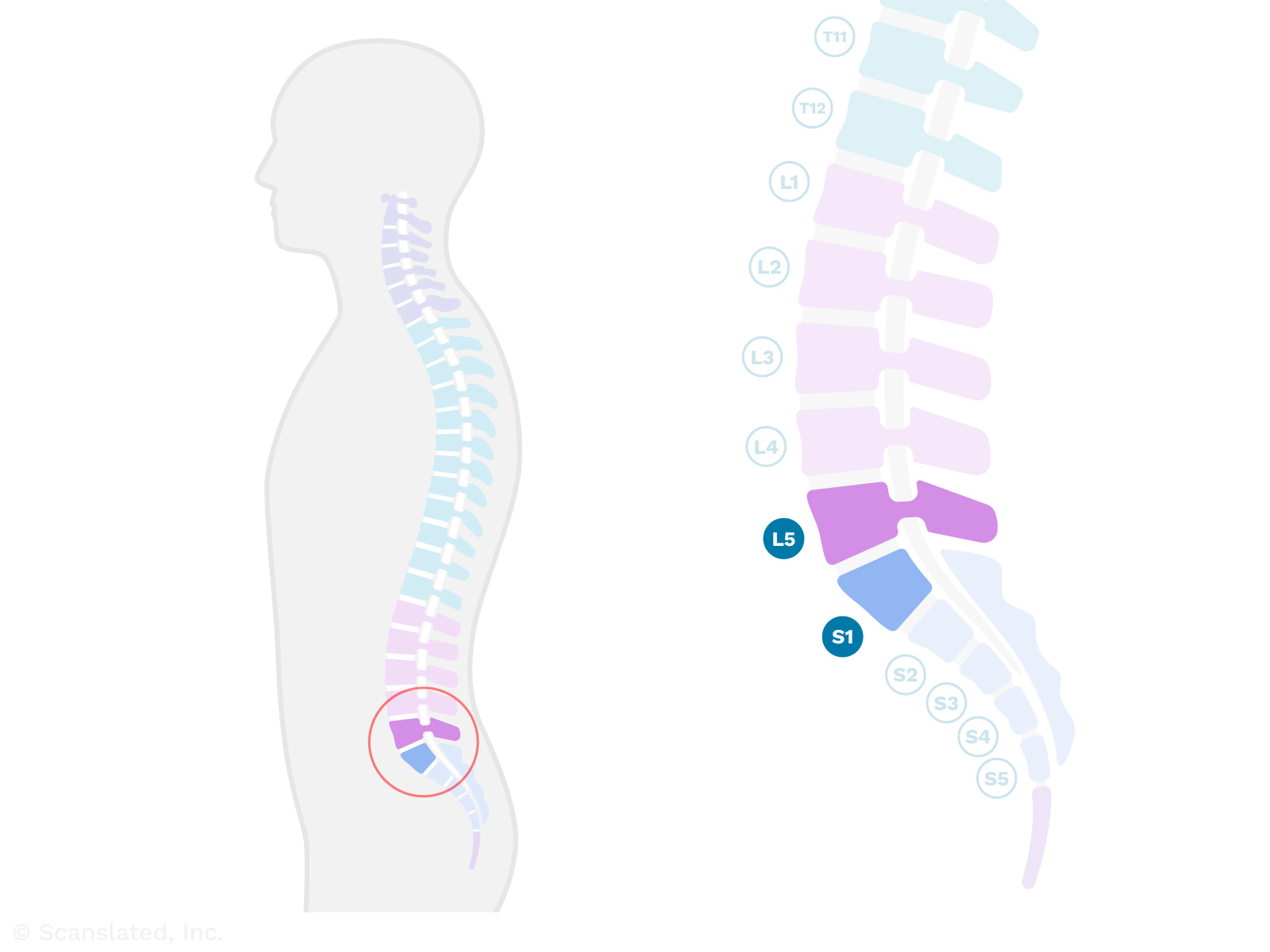
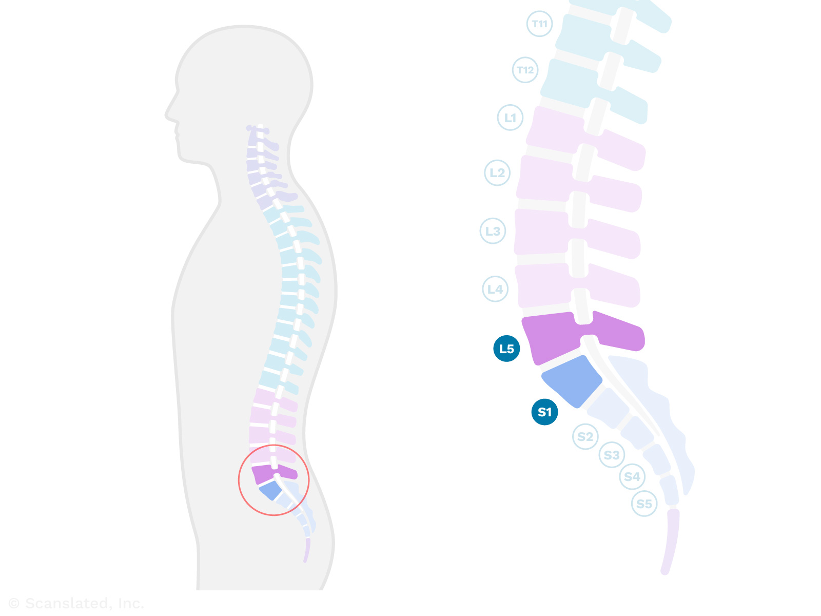
L5-S1
Where the last lumbar vertebra (L5) meets the top of the sacrum (S1).
The sacrum is the large, triangle-shaped “sitting bone” that makes up the back part of the pelvis and is connected to the lower (lumbar) spine. It’s made up of five bones, which usually fuse (join together) during early adulthood.
L5-S1
L5-S1
जहां अंतिम कटि कशेरुका (L5) त्रिकास्थि (S1) के शीर्ष से मिलती है।
त्रिकास्थि बड़ी, त्रिकोण के आकार की “बैठने वाली हड्डी” है जो श्रोणि के पीछे का हिस्सा बनाती है और निचली (काठ) रीढ़ से जुड़ी होती है। यह पाँच हड्डियों से बनी होती है, जो आमतौर पर प्रारंभिक वयस्कता के दौरान जुड़ जाती हैं (एक साथ जुड़ जाती हैं)।
L5-S1
L5-S1
El espacio entre el último hueso lumbar (L5) y el primer hueso sacro (S1). Hay un disco (cojín amortiguador blando) entre los dos huesos y las articulaciones a lo largo de la parte posterior que une los huesos. El nivel L5-S1 se encuentra en la parte más baja de la espalda, justo por encima de los huesos del asiento.
L5-S1
L5-S1
最后一节腰椎(L5)与骶骨顶部(S1)的交接处。
骶骨是一块三角形的大”坐骨”, 构成骨盆的后部, 与下部(腰椎)脊柱相连。它由五块骨头组成, 通常在成年早期会融合(连接)在一起。
L5-S1
L5-S1
Point de rencontre entre la dernière vertèbre lombaire (L5) et la partie supérieure du sacrum (S1).
Le sacrum est le grand « os de l’assise » en forme de triangle qui constitue la partie arrière du bassin et est relié à la colonne vertébrale inférieure (lombaire). Il est composé de cinq os, qui fusionnent généralement au début de l’âge adulte.
suspicious
Possibly abnormal and/or worrisome
suspicious
संदेहास्पद
संभवतः असामान्य और/या चिंताजनक
suspicious
sospechoso
Sospechoso significa la observación de algo en la exploración que es preocupante o que definitivamente no es normal
suspicious
可疑的
可能异常和/或令人担忧
suspicious
suspicieux
Possiblement anormal et/ou inquiétant
marrow
The soft, spongy material inside the bones. This is where blood cells are produced.
Bone marrow plays an important role in the immune system by controlling the number and type of white blood cells in the body. It also maintains the other elements of blood such as red blood cells and platelets.
lesion
An abnormal area, spot, growth, or cyst
lesion
घाव
कोई असामान्य क्षेत्र, धब्बा, वृद्धि या पुटी
lesion
lesión
una zona, mancha o crecimiento anormal
lesion
病灶
异常区域、斑点、赘生物或囊肿
lesion
lésion
Région, point, excroissance ou kyste anormal
Conus Medullaris
The lower end of the spinal cord, which is shaped like a cone or funnel.
The spinal cord usually ends near the top of the lower back, but this can vary from person to person.
Beyond this point, the nerves to the legs continue down through the spinal canal, but there is no longer a solid spinal cord.
Conus Medullaris
कोनस मेडुलैरिस
रीढ़ की हड्डी का निचला सिरा, जो शंकु या कीप जैसा आकार का होता है।
रीढ़ की हड्डी आमतौर पर पीठ के निचले हिस्से के शीर्ष के पास समाप्त होती है, लेकिन यह हर व्यक्ति में अलग-अलग हो सकती है।
इस बिंदु से आगे, पैरों की तंत्रिकाएं रीढ़ की हड्डी की नली से होकर नीचे तक जाती रहती हैं, लेकिन वहां ठोस रीढ़ की हड्डी नहीं रहती।
Conus Medullaris
cono medular
La parte inferior de la médula espinal tiene forma de cono o de embudo. El fin de la medula espinal, suele estar al nivel L1 o L2 de la columna vertebral, pero puede variar entre las personas.
Conus Medullaris
脊髓圆锥
脊髓的下端, 形状像一个圆锥或漏斗。
脊髓的末端通常在下背部顶端, 但也会因人而异。
过了这一点, 通往腿部的神经继续沿椎管下行, 但不再有坚实的脊髓。
Conus Medullaris
cône médullaire
Extrémité inférieure de la moelle épinière en forme de cône ou d’entonnoir.
La moelle épinière se termine généralement près de la partie supérieure du bas du dos, mais cela peut varier d’une personne à l’autre.
Au-delà de ce point, les nerfs des jambes continuent à descendre dans le canal rachidien, mais il n’y a plus de moelle épinière à proprement parler.
Terminates
Ends, stops, or is no longer seen beyond this point
Terminates
समाप्त
इस बिंदु से आगे समाप्त हो जाता है, रुक जाता है, या दिखाई नहीं देता
Terminates
termina
se termina, se detiene, no se ve después de este punto
Terminates
终止
结束、停止或在此点之后不再出现
Terminates
se termine
Se termine, s’arrête ou n’est plus vu au-delà de ce point
level
This term is usually used by radiologists to describe the location of something height-wise in the body (i.e., from head to toe).
For example, if something is “at the L3 level,” that means it’s located in the area of the lower back where the third lumbar vertebra (spine bone) is.
level
स्तर
इस शब्द का प्रयोग आमतौर पर रेडियोलॉजिस्ट द्वारा शरीर में ऊंचाई के अनुसार किसी वस्तु के स्थान (अर्थात सिर से पैर तक) का वर्णन करने के लिए किया जाता है।
उदाहरण के लिए, यदि कोई चीज “एल3 स्तर पर” है, तो इसका मतलब है कि यह पीठ के निचले हिस्से में स्थित है, जहां तीसरी कटि कशेरुका (रीढ़ की हड्डी) होती है।
level
nivel
Normalmente utilizado por los radiólogos para describir la localización de un hallazgo. Es decir, el lugar donde se localiza algo de la cabeza a los pies.
level
水平
放射科医生通常用这个术语来描述某物在人体内(即从头到脚趾)的高度位置。
例如, 如果某物”在 L3 水平”, 这是指它位于下背部区域第三腰椎(椎骨)所在的位置。
level
niveau
Ce terme est généralement utilisé par les radiologues pour décrire l’emplacement de quelque chose dans le corps (c’est-à-dire de la tête aux pieds).
Par exemple, si quelque chose se trouve « au niveau L3 », cela signifie qu’il est situé dans la région du bas du dos où se trouve la troisième vertèbre lombaire (os de la colonne vertébrale).
Normal signal
In MRI, “signal” is the response of the body’s tissues to the magnetic field created by the MRI machine.
Different tissues (bone, muscle, fat, etc.) have different amounts of signal depending on their densities. These differences in signal make the tissues appear darker or brighter on the MRI images. This allows the MRI machine’s computer to build a picture of the inside of the body.
Normal signal means that the body tissue looks as expected on the MRI images.
Normal signal
señal normal
En la IRM, la señal es la reacción de los tejidos del cuerpo al campo magnético creado por la máquina de IRM. Distintos tejidos (hueso, músculo, grasa, etc.) emiten señales diferentes, lo cual permite que la computadora de la máquina de resonancia magnética distinga entre ellos, y así pueda construir una imagen del interior del cuerpo. Una señal normal significa que el tejido del cuerpo tiene el aspecto esperado.
Disc
Spinal discs (or “intervertebral discs”) are the soft, shock-absorbing cushions between spine bones (vertebrae) that allow the spine to flex.
Disc
डिस्क
स्पाइनल डिस्क रीढ़ की हड्डियों के बीच नरम, आघात-अवशोषित कुशन होते हैं जो रीढ़ को लचीला होने देते हैं।
Disc
disco
Los discos vertebrales son el material blando que amortigua los impactos entre los huesos de la columna vertebral.
Disc
椎间盘
椎间盘是椎骨之间的减震软垫, 使脊柱能够弯曲。
Disc
disque
Les disques vertébraux (ou « disques intervértébraux ») sont des coussins souples qui absorbent les chocs entre les os de la colonne vertébrale (les vertèbres) et qui permettent à cette dernière de fléchir.
Levels
This term is usually used by radiologists to describe the location of something height-wise in the body (i.e., from head to toe).
For example, if something is “at the L3 and L4 levels,” that means it’s located in the area of the lower back where the third and fourth lumbar vertebrae (spine bones) are.
Levels
लेवल्स
इस शब्द का प्रयोग आमतौर पर रेडियोलॉजिस्ट द्वारा शरीर में ऊंचाई के अनुसार किसी वस्तु के स्थान (अर्थात सिर से पैर तक) का वर्णन करने के लिए किया जाता है।
उदाहरण के लिए, यदि कोई चीज़ “एल3 और एल4 स्तर पर” है, तो इसका मतलब है कि यह पीठ के निचले हिस्से के क्षेत्र में स्थित है जहां तीसरी और चौथी काठ कशेरुका (रीढ़ की हड्डियां) हैं।
Levels
niveles
Normalmente se utiliza para describir la localización de algo, donde se localiza algo de la cabeza a los pies. Por ejemplo, los niveles de la columna vertebral. Cada hueso de la columna vertebral (vértebra) recibe un número y una letra para designar dónde se encuentra a lo largo de la columna de 33 huesos del cuello / la espalda. Por ejemplo, el tercer hueso de la espalda (lumbar) se denomina L3. Esta parte de la espalda se llama el nivel L3.
Levels
水平
放射科医生通常用这个术语来描述某物在人体内(即从头到脚趾)的高度位置。
例如, 如果某物”在 L3 和 L4 水平”, 这是指它位于下背部区域第三和第四腰椎(椎骨)所在的位置。
Levels
niveaux
Ce terme est généralement utilisé par les radiologues pour décrire l’emplacement de quelque chose dans le corps (c’est-à-dire de la tête aux pieds).
Par exemple, si quelque chose se trouve « au niveau L3 et L4 », cela signifie qu’il est situé dans la région du bas du dos où se trouve la troisième et la quatrième vertèbre lombaire (os de la colonne vertébrale).
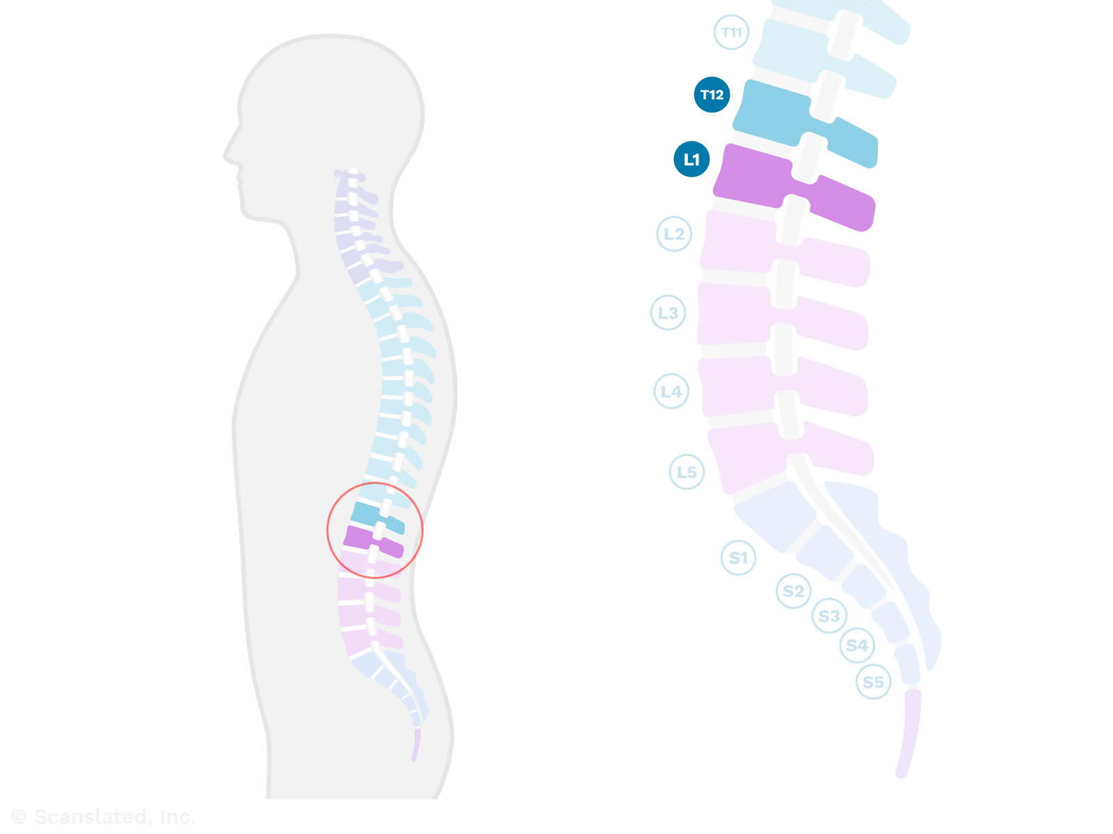
T12-L1
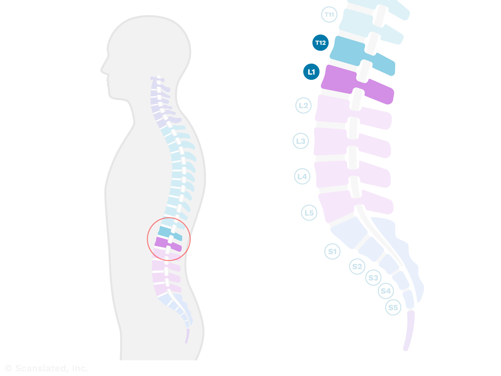
Where the last thoracic vertebra (T12) meets the first lumbar vertebra (L1). This is where the upper/mid back meets the lower back (below the rib cage).
T12-L1
T12-L1
जहां अंतिम वक्षीय कशेरुका (T12) प्रथम कटि कशेरुका (L1) से मिलती है। यह वह जगह है जहां ऊपरी/मध्य पीठ निचली पीठ (पसलियों के पिंजरे के नीचे) से मिलती है।
T12-L1
T12-L1
El espacio entre los cuerpos vertebrales torácicos más bajos y la primera vértebra lumbar (huesos de la espalda), donde la parte media de la espalda se encuentra con la parte baja. Hay un disco (cojín amortiguador blando) entre los dos huesos y las articulaciones a lo largo de la parte posterior que une a los huesos.
T12-L1
T12-L1
最后一节胸椎(T12)与第一节腰椎(L1)的交接处。这是上/中背部与下背部(肋骨下方)的交汇处。
T12-L1
T12-L1
Point de rencontre entre la dernière vertèbre thoracique (T12) et la première vertèbre lombaire (L1). C’est l’endroit où le haut et le milieu du dos rencontrent le bas du dos (sous la cage thoracique).
disc
Spinal discs (or “intervertebral discs”) are the soft, shock-absorbing cushions between spine bones (vertebrae) that allow the spine to flex.
disc
डिस्क
स्पाइनल डिस्क रीढ़ की हड्डियों के बीच नरम, आघात-अवशोषित कुशन होते हैं जो रीढ़ को लचीला होने देते हैं।
disc
disco
Los discos vertebrales son el material blando que amortigua los impactos entre los huesos de la columna vertebral.
disc
椎间盘
椎间盘是椎骨之间的减震软垫, 使脊柱能够弯曲。
disc
disque
Les disques vertébraux (ou « disques intervértébraux ») sont des coussins souples qui absorbent les chocs entre les os de la colonne vertébrale (les vertèbres) et qui permettent à cette dernière de fléchir.
spinal canal stenosis
Narrowing of the spinal canal (the space where the spinal cord and nerves travel through the spine bones). This can put pressure on the spinal cord and nerves, which can lead to symptoms like pain, numbness, or weakness.
Stenosis (narrowing) can be caused by a bulging disc, bone spurs, or enlargement of the ligaments in the back of the spine.
neural foraminal narrowing
Narrowing (stenosis) of the neural foramina, which can be caused by bone spurs or bulging discs.
A “foramen” (plural: “foramina”) is a normal hole or tunnel in a bone.
In the spine, neural foramina are openings on either side of the vertebrae (spine bones) where the nerves travel. Nerves exit the spinal canal through these openings at each level of the spine, going out to the left and right sides of the body.
Narrowing of these openings can sometimes be a cause for pain.
neural foraminal narrowing
तंत्रिका छिद्र का संकुचन
तंत्रिका छिद्रों का संकुचित होना (स्टेनोसिस), जो अस्थि स्पर्स या उभरी हुई डिस्क के कारण हो सकता है।
एक “फोरैमन” (बहुवचन: “फोरैमिना”) हड्डी में एक सामान्य छेद या सुरंग है।
रीढ़ में, न्यूरल फोरैमिना कशेरुकाओं (रीढ़ की हड्डियों) के दोनों ओर खुले होते हैं जहां तंत्रिकाएं यात्रा करती हैं। रीढ़ की हड्डी के प्रत्येक स्तर पर इन छिद्रों के माध्यम से नसें रीढ़ की हड्डी की नहर से बाहर निकलती हैं, शरीर के बाएँ और दाएँ तरफ जाती हैं।
इन छिद्रों का संकुचित होना कभी-कभी दर्द का कारण बन सकता है।
neural foraminal narrowing
pinzamiento del agujero intervertebral
Estrechez de las aberturas en cada lado de los huesos de la columna vertebral por donde recorren los nervios. Los nervios salen del canal espinal por cada nivel de la columna vertebral y transmiten señales de movimiento y de sensación a las diferentes partes del cuerpo. A cada nivel, hay un nervio que se dirige al lado derecho, y un nervio que se dirige al lado izquierdo del cuerpo. Estos nervios recorren por el agujero intervertebral en cada lado al salir de la columna vertebral. El estrechamiento de estas aberturas puede ser causado por espolones óseos o hernias discales.
neural foraminal narrowing
神经孔狭窄
神经孔变窄(狭窄), 可能由骨刺或椎间盘膨出引起。
骨骼中存在正常的孔洞或隧道, 称之为”孔”。
在脊柱中, 神经孔是脊柱骨(椎骨)一侧的有神经穿过的开口。神经通过每个脊柱水平的这些开口离开椎管, 通向身体的左右两侧。
这些开口变窄有时会导致疼痛。
neural foraminal narrowing
rétrécissement du foramen neural
Le rétrécissement (sténose) du foramen neural peut être causé par des épines osseuses ou des hernies discales.
Le « foramen » (pluriel : « foramina » ) est un trou ou un tunnel normal dans un os.
Dans la colonne vertébrale, les foramina neuraux sont des ouvertures de chaque côté des vertèbres (les os de la colonne vertébrale) où passent les nerfs. Les nerfs sortent du canal rachidien par ces ouvertures à chaque niveau de la colonne vertébrale et partent à droite et à gauche du corps.
Le rétrécissement de ces ouvertures peut parfois provoquer de la douleur.
L1-L2
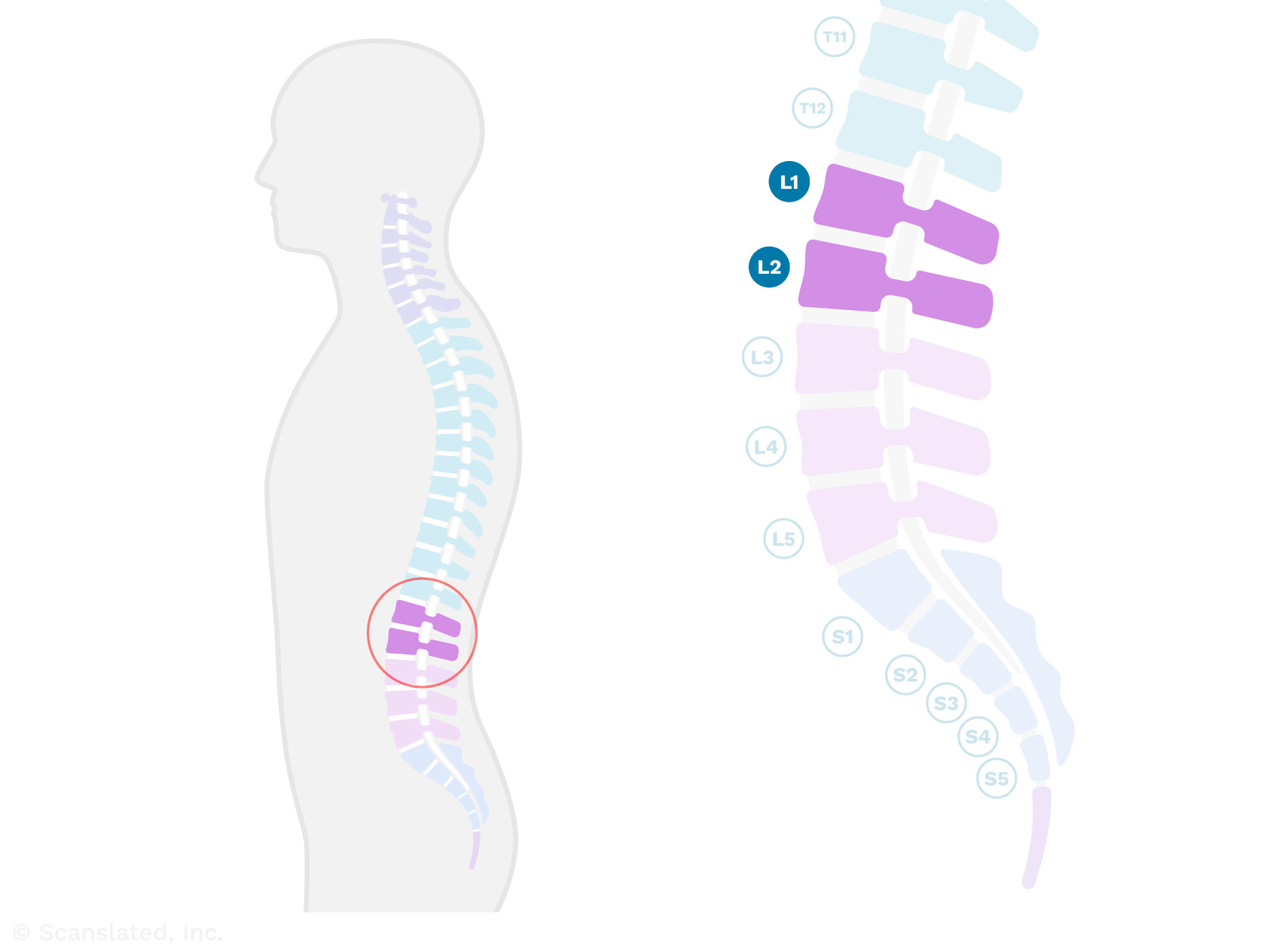
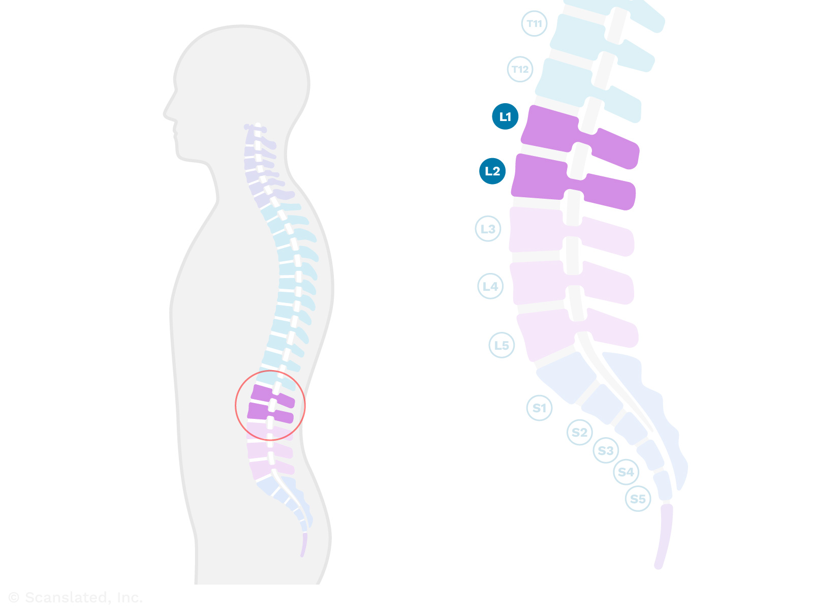
At the level of the 1st and 2nd lumbar vertebrae (spine bones) in the lower back.
There are usually five bones in the lumbar spine, which begins just below the rib cage and continues down through the lower back.
L1-L2
L1-L2
पीठ के निचले हिस्से में प्रथम और द्वितीय काठ कशेरुका (रीढ़ की हड्डी) के स्तर पर।
काठ की रीढ़ में आमतौर पर पांच हड्डियां होती हैं, जो पसलियों के पिंजरे के ठीक नीचे से शुरू होती हैं और पीठ के निचले हिस्से से नीचे तक जाती हैं।
L1-L2
L1-L2
Entre los cuerpos vertebrales lumbares 1º y 2º (huesos de la espalda baja). Hay un disco (cojín amortiguador blando) entre los dos huesos y las articulaciones a lo largo de la parte posterior que une los huesos.
L1-L2
L1-L2
位于下背部第1和第2腰椎(脊椎骨)的位置。
腰椎通常有五块骨头, 从肋骨下方开始, 一直延伸到下背部。
L1-L2
L1-L2
Au niveau des 1ère et 2ème vertèbres lombaires (os de la colonne vertébrale) dans le bas du dos.
La colonne vertébrale lombaire, qui commence juste en dessous de la cage thoracique et se poursuit jusqu’au bas du dos, compte généralement cinq os.
disc
Spinal discs (or “intervertebral discs”) are the soft, shock-absorbing cushions between spine bones (vertebrae) that allow the spine to flex.
disc
डिस्क
स्पाइनल डिस्क रीढ़ की हड्डियों के बीच नरम, आघात-अवशोषित कुशन होते हैं जो रीढ़ को लचीला होने देते हैं।
disc
disco
Los discos vertebrales son el material blando que amortigua los impactos entre los huesos de la columna vertebral.
disc
椎间盘
椎间盘是椎骨之间的减震软垫, 使脊柱能够弯曲。
disc
disque
Les disques vertébraux (ou « disques intervértébraux ») sont des coussins souples qui absorbent les chocs entre les os de la colonne vertébrale (les vertèbres) et qui permettent à cette dernière de fléchir.
spinal canal stenosis
Narrowing of the spinal canal (the space where the spinal cord and nerves travel through the spine bones). This can put pressure on the spinal cord and nerves, which can lead to symptoms like pain, numbness, or weakness.
Stenosis (narrowing) can be caused by a bulging disc, bone spurs, or enlargement of the ligaments in the back of the spine.
neural foraminal narrowing
Narrowing (stenosis) of the neural foramina, which can be caused by bone spurs or bulging discs.
A “foramen” (plural: “foramina”) is a normal hole or tunnel in a bone.
In the spine, neural foramina are openings on either side of the vertebrae (spine bones) where the nerves travel. Nerves exit the spinal canal through these openings at each level of the spine, going out to the left and right sides of the body.
Narrowing of these openings can sometimes be a cause for pain.
neural foraminal narrowing
तंत्रिका छिद्र का संकुचन
तंत्रिका छिद्रों का संकुचित होना (स्टेनोसिस), जो अस्थि स्पर्स या उभरी हुई डिस्क के कारण हो सकता है।
एक “फोरैमन” (बहुवचन: “फोरैमिना”) हड्डी में एक सामान्य छेद या सुरंग है।
रीढ़ में, न्यूरल फोरैमिना कशेरुकाओं (रीढ़ की हड्डियों) के दोनों ओर खुले होते हैं जहां तंत्रिकाएं यात्रा करती हैं। रीढ़ की हड्डी के प्रत्येक स्तर पर इन छिद्रों के माध्यम से नसें रीढ़ की हड्डी की नहर से बाहर निकलती हैं, शरीर के बाएँ और दाएँ तरफ जाती हैं।
इन छिद्रों का संकुचित होना कभी-कभी दर्द का कारण बन सकता है।
neural foraminal narrowing
pinzamiento del agujero intervertebral
Estrechez de las aberturas en cada lado de los huesos de la columna vertebral por donde recorren los nervios. Los nervios salen del canal espinal por cada nivel de la columna vertebral y transmiten señales de movimiento y de sensación a las diferentes partes del cuerpo. A cada nivel, hay un nervio que se dirige al lado derecho, y un nervio que se dirige al lado izquierdo del cuerpo. Estos nervios recorren por el agujero intervertebral en cada lado al salir de la columna vertebral. El estrechamiento de estas aberturas puede ser causado por espolones óseos o hernias discales.
neural foraminal narrowing
神经孔狭窄
神经孔变窄(狭窄), 可能由骨刺或椎间盘膨出引起。
骨骼中存在正常的孔洞或隧道, 称之为”孔”。
在脊柱中, 神经孔是脊柱骨(椎骨)一侧的有神经穿过的开口。神经通过每个脊柱水平的这些开口离开椎管, 通向身体的左右两侧。
这些开口变窄有时会导致疼痛。
neural foraminal narrowing
rétrécissement du foramen neural
Le rétrécissement (sténose) du foramen neural peut être causé par des épines osseuses ou des hernies discales.
Le « foramen » (pluriel : « foramina » ) est un trou ou un tunnel normal dans un os.
Dans la colonne vertébrale, les foramina neuraux sont des ouvertures de chaque côté des vertèbres (les os de la colonne vertébrale) où passent les nerfs. Les nerfs sortent du canal rachidien par ces ouvertures à chaque niveau de la colonne vertébrale et partent à droite et à gauche du corps.
Le rétrécissement de ces ouvertures peut parfois provoquer de la douleur.
L2-L3
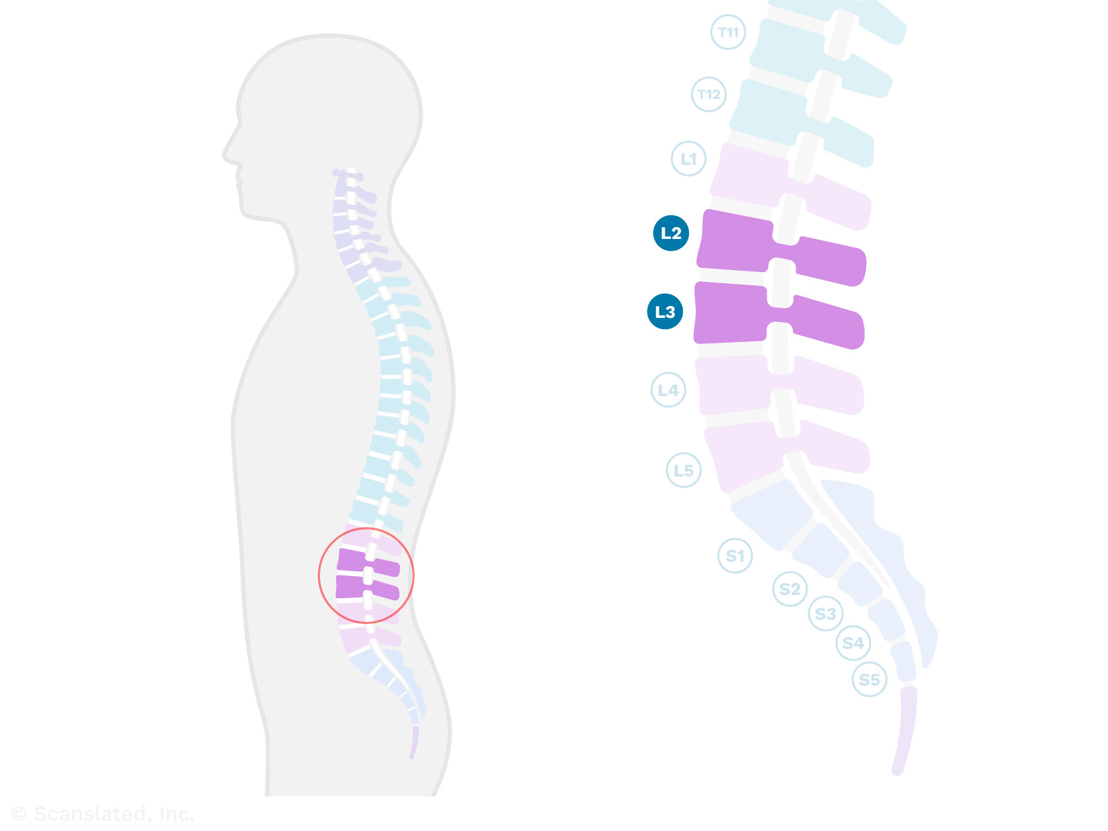
At the level of the 2nd and 3rd lumbar vertebrae (spine bones) in the lower back.
There are usually five bones in the lumbar spine, which begins just below the rib cage and continues down through the lower back.
L2-L3
L2-L3
पीठ के निचले हिस्से में दूसरे और तीसरे काठ कशेरुका (रीढ़ की हड्डी) के स्तर पर।
काठ की रीढ़ में आमतौर पर पांच हड्डियां होती हैं, जो पसलियों के पिंजरे के ठीक नीचे से शुरू होती हैं और पीठ के निचले हिस्से से नीचे तक जाती हैं।
L2-L3
L2-L3
Entre los cuerpos vertebrales lumbares 2º y 3º (huesos de la parte baja de la espalda). Hay un disco (cojín amortiguador blando) entre los dos huesos y las articulaciones a lo largo de la parte posterior que une los huesos.
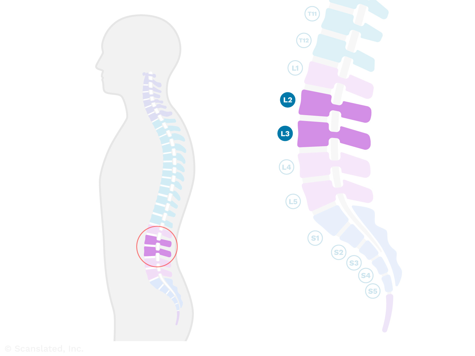
L2-L3
L2-L3
位于下背部第2和第3腰椎(脊椎骨)的位置。
腰椎通常有五块骨头, 从肋骨下方开始, 一直延伸到下背部。
L2-L3
L2-L3
Au niveau de la 2ème à la 3ème vertèbre lombaire (os de la colonne vertébrale) dans le bas du dos.
La colonne vertébrale lombaire, qui commence juste en dessous de la cage thoracique et se poursuit jusqu’au bas du dos, compte généralement cinq os.
disc desiccation
When a spinal disc (soft cushion between spine bones) loses some of its water content and dries out. This happens over time with wear and tear.
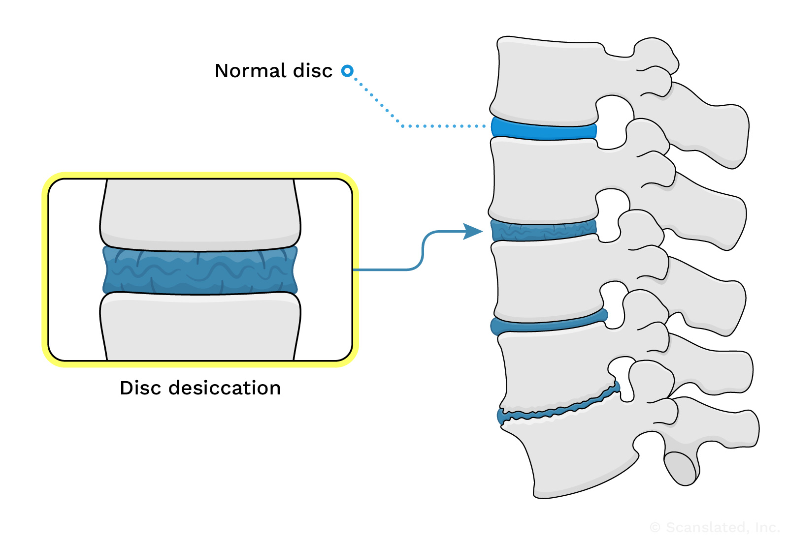
disc
Spinal discs (or “intervertebral discs”) are the soft, shock-absorbing cushions between spine bones (vertebrae) that allow the spine to flex.
disc
डिस्क
स्पाइनल डिस्क रीढ़ की हड्डियों के बीच नरम, आघात-अवशोषित कुशन होते हैं जो रीढ़ को लचीला होने देते हैं।
disc
disco
Los discos vertebrales son el material blando que amortigua los impactos entre los huesos de la columna vertebral.
disc
椎间盘
椎间盘是椎骨之间的减震软垫, 使脊柱能够弯曲。
disc
disque
Les disques vertébraux (ou « disques intervértébraux ») sont des coussins souples qui absorbent les chocs entre les os de la colonne vertébrale (les vertèbres) et qui permettent à cette dernière de fléchir.
spinal canal stenosis
Narrowing of the spinal canal (the space where the spinal cord and nerves travel through the spine bones). This can put pressure on the spinal cord and nerves, which can lead to symptoms like pain, numbness, or weakness.
Stenosis (narrowing) can be caused by a bulging disc, bone spurs, or enlargement of the ligaments in the back of the spine.
neural foraminal narrowing
Narrowing (stenosis) of the neural foramina, which can be caused by bone spurs or bulging discs.
A “foramen” (plural: “foramina”) is a normal hole or tunnel in a bone.
In the spine, neural foramina are openings on either side of the vertebrae (spine bones) where the nerves travel. Nerves exit the spinal canal through these openings at each level of the spine, going out to the left and right sides of the body.
Narrowing of these openings can sometimes be a cause for pain.
neural foraminal narrowing
तंत्रिका छिद्र का संकुचन
तंत्रिका छिद्रों का संकुचित होना (स्टेनोसिस), जो अस्थि स्पर्स या उभरी हुई डिस्क के कारण हो सकता है।
एक “फोरैमन” (बहुवचन: “फोरैमिना”) हड्डी में एक सामान्य छेद या सुरंग है।
रीढ़ में, न्यूरल फोरैमिना कशेरुकाओं (रीढ़ की हड्डियों) के दोनों ओर खुले होते हैं जहां तंत्रिकाएं यात्रा करती हैं। रीढ़ की हड्डी के प्रत्येक स्तर पर इन छिद्रों के माध्यम से नसें रीढ़ की हड्डी की नहर से बाहर निकलती हैं, शरीर के बाएँ और दाएँ तरफ जाती हैं।
इन छिद्रों का संकुचित होना कभी-कभी दर्द का कारण बन सकता है।
neural foraminal narrowing
pinzamiento del agujero intervertebral
Estrechez de las aberturas en cada lado de los huesos de la columna vertebral por donde recorren los nervios. Los nervios salen del canal espinal por cada nivel de la columna vertebral y transmiten señales de movimiento y de sensación a las diferentes partes del cuerpo. A cada nivel, hay un nervio que se dirige al lado derecho, y un nervio que se dirige al lado izquierdo del cuerpo. Estos nervios recorren por el agujero intervertebral en cada lado al salir de la columna vertebral. El estrechamiento de estas aberturas puede ser causado por espolones óseos o hernias discales.
neural foraminal narrowing
神经孔狭窄
神经孔变窄(狭窄), 可能由骨刺或椎间盘膨出引起。
骨骼中存在正常的孔洞或隧道, 称之为”孔”。
在脊柱中, 神经孔是脊柱骨(椎骨)一侧的有神经穿过的开口。神经通过每个脊柱水平的这些开口离开椎管, 通向身体的左右两侧。
这些开口变窄有时会导致疼痛。
neural foraminal narrowing
rétrécissement du foramen neural
Le rétrécissement (sténose) du foramen neural peut être causé par des épines osseuses ou des hernies discales.
Le « foramen » (pluriel : « foramina » ) est un trou ou un tunnel normal dans un os.
Dans la colonne vertébrale, les foramina neuraux sont des ouvertures de chaque côté des vertèbres (les os de la colonne vertébrale) où passent les nerfs. Les nerfs sortent du canal rachidien par ces ouvertures à chaque niveau de la colonne vertébrale et partent à droite et à gauche du corps.
Le rétrécissement de ces ouvertures peut parfois provoquer de la douleur.
L3-L4
At the level of the 3rd and 4th lumbar vertebrae (spine bones) in the lower back.
There are usually five bones in the lumbar spine, which begins just below the rib cage and continues down through the lower back.
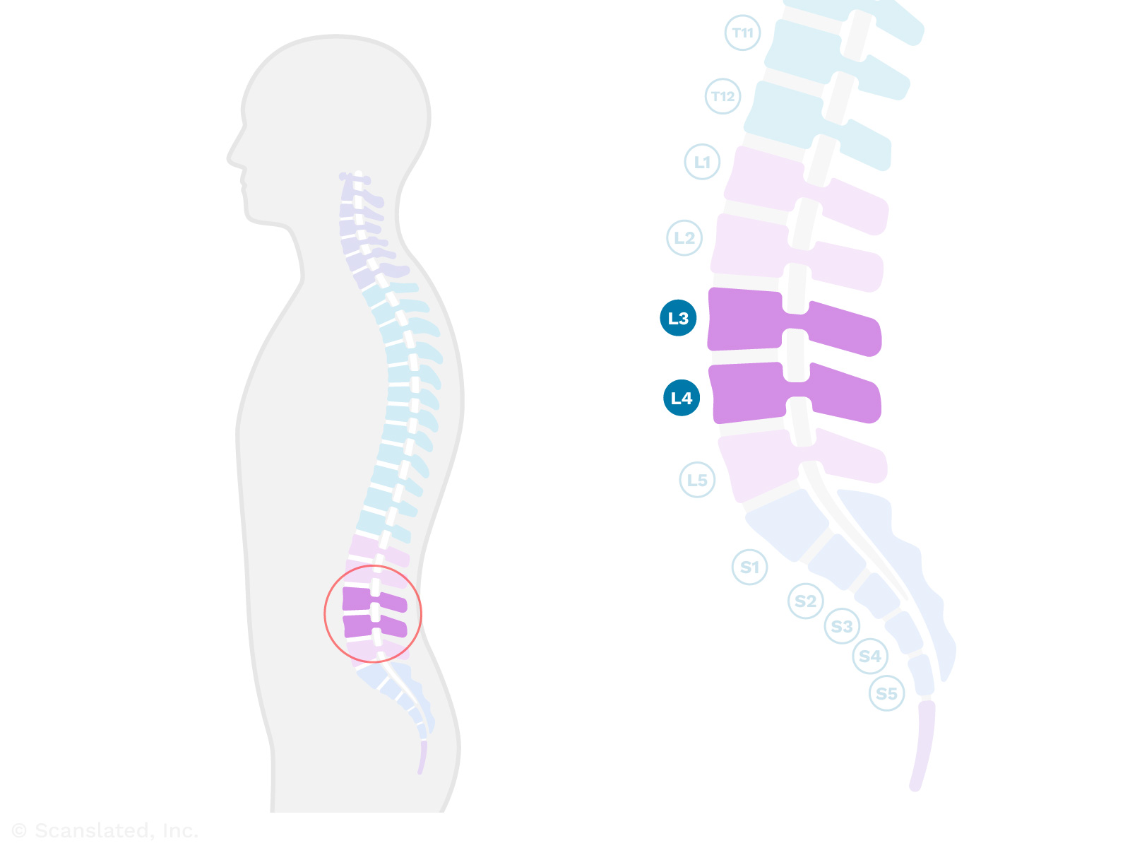
L3-L4
L3-L4
पीठ के निचले हिस्से में तीसरी और चौथी काठ कशेरुका (रीढ़ की हड्डियाँ) के स्तर पर।
काठ की रीढ़ में आमतौर पर पांच हड्डियां होती हैं, जो पसलियों के पिंजरे के ठीक नीचे से शुरू होती हैं और पीठ के निचले हिस्से से नीचे तक जाती हैं।
L3-L4
L3-L4
Entre la 3ª y 4ª vértebra lumbar (huesos de la parte baja de la espalda). Hay un disco (cojín amortiguador blando) entre los dos huesos y las articulaciones a lo largo de la parte posterior que une los huesos.
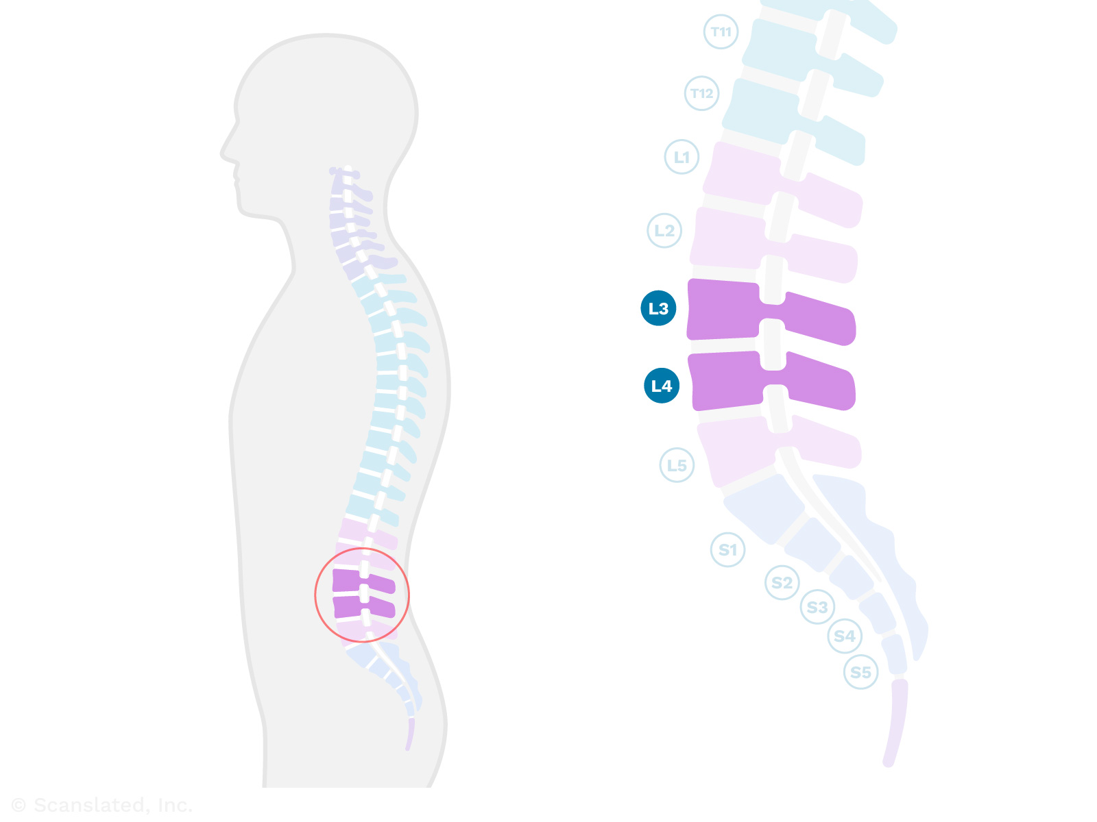
L3-L4
L3-L4
位于下腰部第3和第4腰椎(脊椎骨)的位置。
腰椎通常有五块骨头, 从肋骨下方开始, 一直延伸到下背部。
L3-L4
L3-L4
Au niveau des 3ème et 4ème vertèbres lombaires (os de la colonne vertébrale) dans le bas du dos.
La colonne vertébrale lombaire, qui commence juste en dessous de la cage thoracique et se poursuit jusqu’au bas du dos, compte généralement cinq os.
disc bulge
A disc bulge is an outpouching of a spinal disc (the soft cushion between spine bones).
Disc bulges are fairly common, and may not cause any problems. In some cases, larger disc bulges can put pressure on nearby nerves and cause pain or weakness.
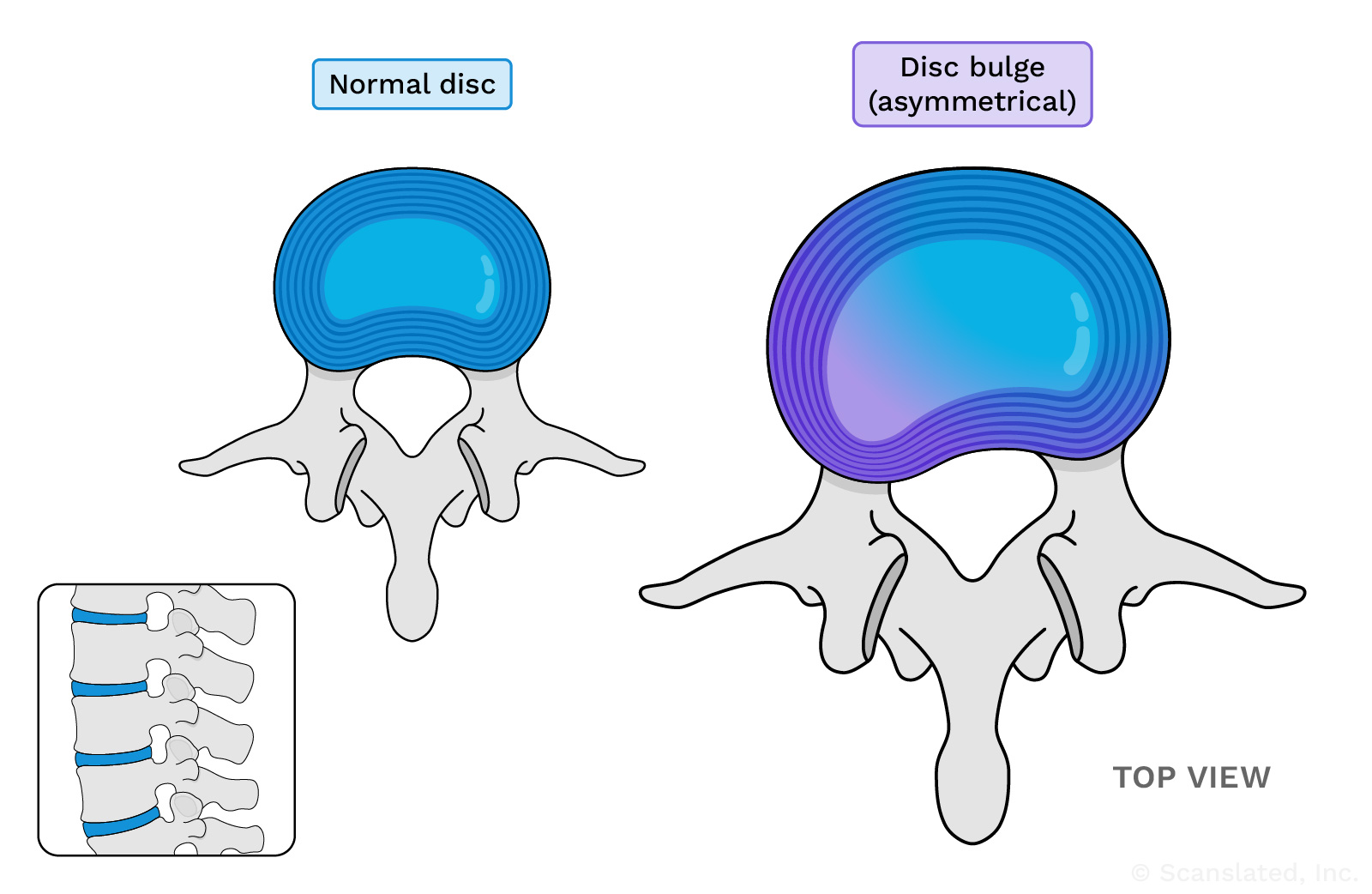
This diagram shows an asymmetrical disc bulge on the left side of the disc as an example. This is not based on your report, as disc bulges can vary in size and location.
disc bulge
डिस्क उभार
डिस्क उभार स्पाइनल डिस्क (रीढ़ की हड्डियों के बीच नरम गद्दी) का एक फैलाव है।
डिस्क का उभार काफी आम है, और इससे कोई समस्या नहीं होती। कुछ मामलों में, डिस्क का बड़ा उभार आस-पास की नसों पर दबाव डाल सकता है और दर्द या कमजोरी पैदा कर सकता है।
This diagram shows an asymmetrical disc bulge on the left side of the disc as an example. This is not based on your report, as disc bulges can vary in size and location.
disc bulge
abombamiento de disco
El abombamiento de disco es una evaginación del material blando del disco entre dos huesos de la columna vertebral. Los abombamientos de disco son bastantes comunes y quizás no causen ningún problema. A veces, los abombamientos de disco más grandes pueden causar dolor o debilidad cuando presionan el espacio por donde discurren la médula espinal y los nervios (el canal espinal), o porque estrechan los túneles por donde salen los nervios en su recorrido hacia el lado derecho e izquierdo del organismo.
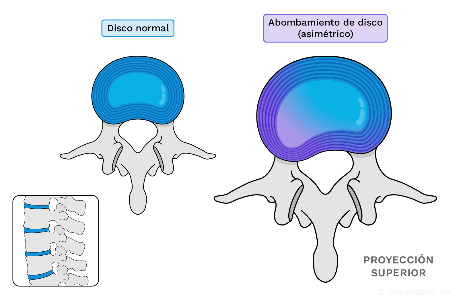
This diagram shows an asymmetrical disc bulge on the left side of the disc as an example. This is not based on your report, as disc bulges can vary in size and location.
disc bulge
椎间盘膨出
椎间盘膨出是指椎间盘(椎骨之间的软骨垫)外扩。
椎间盘膨出相当常见, 可能不会引起任何问题。在某些情况下, 较大范围的椎间盘膨出会对附近的神经造成压迫, 导致疼痛或无力。
This diagram shows an asymmetrical disc bulge on the left side of the disc as an example. This is not based on your report, as disc bulges can vary in size and location.
disc bulge
hernie discale
Une hernie discale est la saillie d’un disque intervertébral (le coussin mou entre les os de la colonne vertébrale).
Les hernies discales sont assez courantes et ne posent généralement pas de problèmes. Dans certains cas, des hernies discales plus importantes peuvent exercer une pression plus importante sur les nefs à proximité et provoquer des douleurs ou de la faiblesse.
This diagram shows an asymmetrical disc bulge on the left side of the disc as an example. This is not based on your report, as disc bulges can vary in size and location.
bilateral facet arthropathy
Wear-and-tear changes of the facet joints, which are the small joints that link the vertebrae (spine bones) together. The facet joints allow the spine to bend forward and backward.
These changes are common with age, and can sometimes be a cause for back or neck pain.
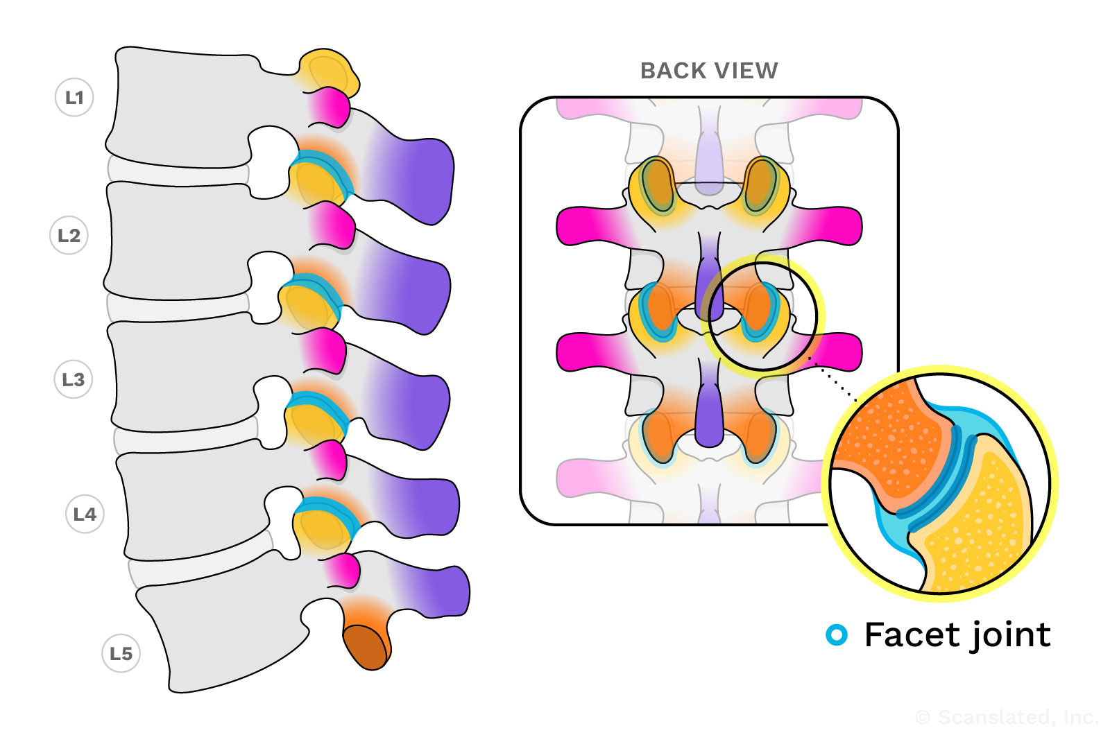
Facet joints are highlighted here in blue. Facet joints are present all along the spine; the lumbar section is shown here as an example.
bilateral facet arthropathy
द्विपक्षीय पहलू आर्थ्रोपैथी
पहलू जोड़ों में टूट-फूट से होने वाले परिवर्तन, जो कि छोटे जोड़ होते हैं जो कशेरुकाओं (रीढ़ की हड्डियों) को एक साथ जोड़ते हैं। पहलू जोड़ रीढ़ को आगे और पीछे झुकने की अनुमति देते हैं।
ये परिवर्तन उम्र के साथ आम हैं और कभी-कभी पीठ या गर्दन में दर्द का कारण भी बन सकते हैं।
Facet joints are highlighted here in blue. Facet joints are present all along the spine; the lumbar section is shown here as an example.
bilateral facet arthropathy
artropatía facetaria bilateral
Cambios por desgaste de las articulaciones pequeñas (facetarias) que unen a los huesos de la columna vertebral (vertebras). Estos cambios son habituales con el envejecimiento. A veces puede ser una causa de dolor de espalda.
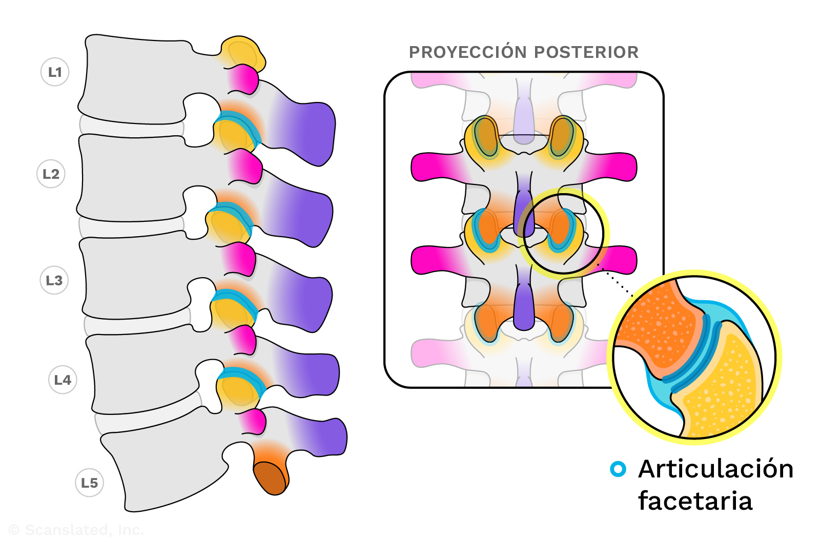
Facet joints are highlighted here in blue. Facet joints are present all along the spine; the lumbar section is shown here as an example.
bilateral facet arthropathy
双侧小关节病
小关节(连接脊柱骨 [椎骨] 的小关节)的磨损性改变。小关节使脊柱可以向前和向后弯曲。
随着年龄的增长, 这种改变很常见, 有时会引起背部或颈部疼痛。
Facet joints are highlighted here in blue. Facet joints are present all along the spine; the lumbar section is shown here as an example.
bilateral facet arthropathy
arthropathie facettaire bilatérale
Usure des facettes vertébrales qui sont de petites articulations reliant les vertèbres (les os de la colonne vertébrale) ensemble. Les facettes vertébrales permettent à la colonne vertébrale de se courber en avant et en arrière.
Ces modifications sont courantes avec l’âge et peuvent parfois être la cause de douleurs dans le dos ou dans le cou.
Facet joints are highlighted here in blue. Facet joints are present all along the spine; the lumbar section is shown here as an example.
No significant
None seen on the scan, or not enough to mention
No significant
कोई महत्वपूर्ण नहीं
स्कैन में कुछ भी नहीं दिखा, या उल्लेख करने लायक भी नहीं
No significant
No significativo
no se ha visto nada en la exploración, o no es lo suficiente para mencionar
No significant
不显著
扫描影像上看不到, 或不足以提及
No significant
pas significatif
Aucun(e) vue(e) sur l’image, pas suffisant pour être mentionné(e)
spinal canal stenosis
Narrowing of the spinal canal (the space where the spinal cord and nerves travel through the spine bones). This can put pressure on the spinal cord and nerves, which can lead to symptoms like pain, numbness, or weakness.
Stenosis (narrowing) can be caused by a bulging disc, bone spurs, or enlargement of the ligaments in the back of the spine.
bilateral
Involving or referring to both the left and right sides
neural foraminal narrowing
Narrowing (stenosis) of the neural foramina, which can be caused by bone spurs or bulging discs.
A “foramen” (plural: “foramina”) is a normal hole or tunnel in a bone.
In the spine, neural foramina are openings on either side of the vertebrae (spine bones) where the nerves travel. Nerves exit the spinal canal through these openings at each level of the spine, going out to the left and right sides of the body.
Narrowing of these openings can sometimes be a cause for pain.
neural foraminal narrowing
तंत्रिका छिद्र का संकुचन
तंत्रिका छिद्रों का संकुचित होना (स्टेनोसिस), जो अस्थि स्पर्स या उभरी हुई डिस्क के कारण हो सकता है।
एक “फोरैमन” (बहुवचन: “फोरैमिना”) हड्डी में एक सामान्य छेद या सुरंग है।
रीढ़ में, न्यूरल फोरैमिना कशेरुकाओं (रीढ़ की हड्डियों) के दोनों ओर खुले होते हैं जहां तंत्रिकाएं यात्रा करती हैं। रीढ़ की हड्डी के प्रत्येक स्तर पर इन छिद्रों के माध्यम से नसें रीढ़ की हड्डी की नहर से बाहर निकलती हैं, शरीर के बाएँ और दाएँ तरफ जाती हैं।
इन छिद्रों का संकुचित होना कभी-कभी दर्द का कारण बन सकता है।
neural foraminal narrowing
pinzamiento del agujero intervertebral
Estrechez de las aberturas en cada lado de los huesos de la columna vertebral por donde recorren los nervios. Los nervios salen del canal espinal por cada nivel de la columna vertebral y transmiten señales de movimiento y de sensación a las diferentes partes del cuerpo. A cada nivel, hay un nervio que se dirige al lado derecho, y un nervio que se dirige al lado izquierdo del cuerpo. Estos nervios recorren por el agujero intervertebral en cada lado al salir de la columna vertebral. El estrechamiento de estas aberturas puede ser causado por espolones óseos o hernias discales.
neural foraminal narrowing
神经孔狭窄
神经孔变窄(狭窄), 可能由骨刺或椎间盘膨出引起。
骨骼中存在正常的孔洞或隧道, 称之为”孔”。
在脊柱中, 神经孔是脊柱骨(椎骨)一侧的有神经穿过的开口。神经通过每个脊柱水平的这些开口离开椎管, 通向身体的左右两侧。
这些开口变窄有时会导致疼痛。
neural foraminal narrowing
rétrécissement du foramen neural
Le rétrécissement (sténose) du foramen neural peut être causé par des épines osseuses ou des hernies discales.
Le « foramen » (pluriel : « foramina » ) est un trou ou un tunnel normal dans un os.
Dans la colonne vertébrale, les foramina neuraux sont des ouvertures de chaque côté des vertèbres (les os de la colonne vertébrale) où passent les nerfs. Les nerfs sortent du canal rachidien par ces ouvertures à chaque niveau de la colonne vertébrale et partent à droite et à gauche du corps.
Le rétrécissement de ces ouvertures peut parfois provoquer de la douleur.
L4-L5
At the level of the 4th and 5th lumbar vertebrae (spine bones) in the lower back.
There are usually five bones in the lumbar spine, which begins just below the rib cage and continues down through the lower back.
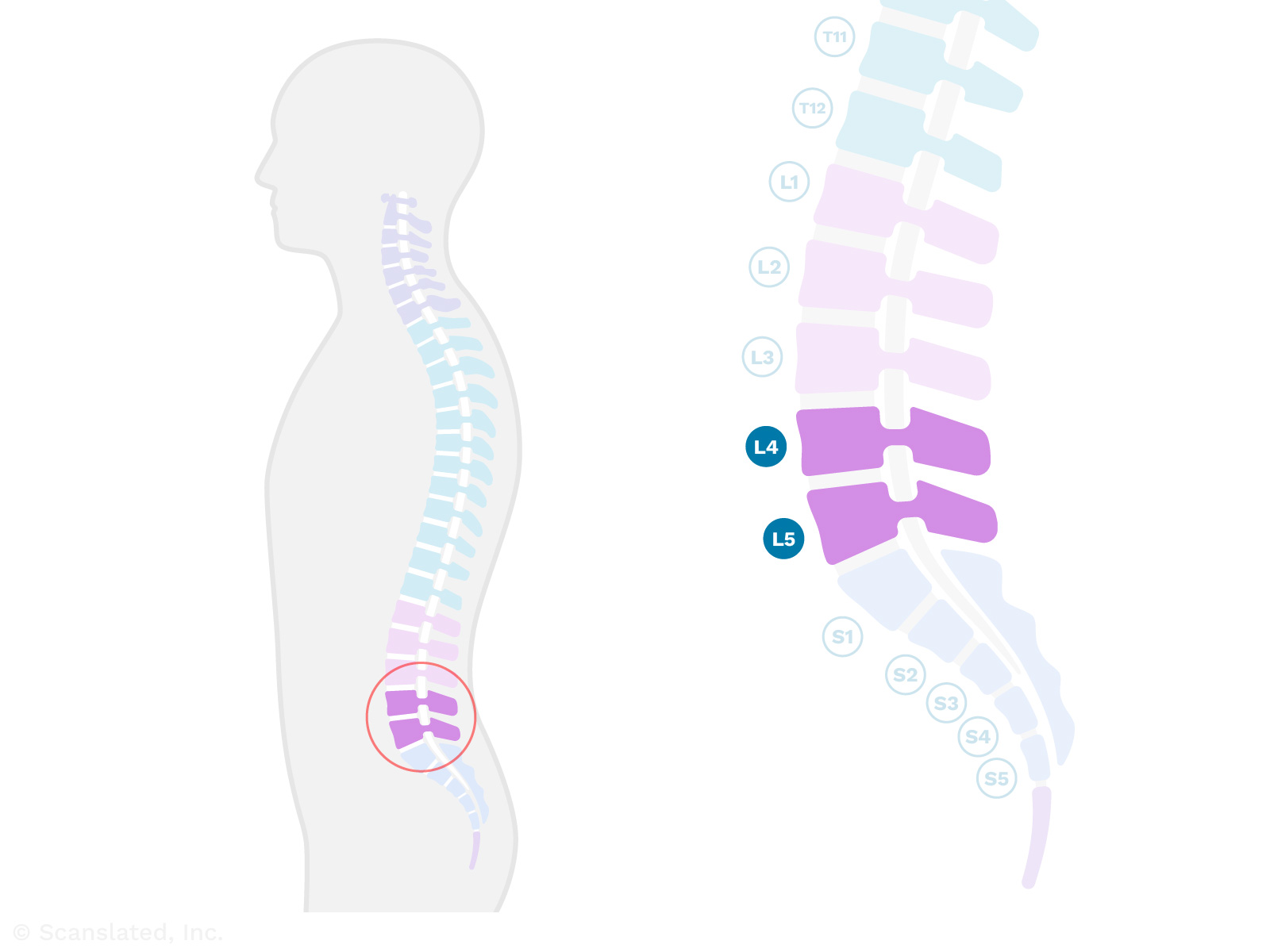
L4-L5
L4-L5
निचली पीठ में चौथी और पांचवीं काठ कशेरुका (रीढ़ की हड्डी) के स्तर पर।
काठ की रीढ़ में आमतौर पर पांच हड्डियां होती हैं, जो पसलियों के पिंजरे के ठीक नीचे से शुरू होती हैं और पीठ के निचले हिस्से से नीचे तक जाती हैं।
L4-L5
L4-L5
Entre los cuerpos vertebrales lumbares 4º y 5º (huesos de la parte baja de la espalda). Hay un disco (cojín amortiguador blando) entre los dos huesos y las articulaciones a lo largo de la parte posterior que une los huesos.
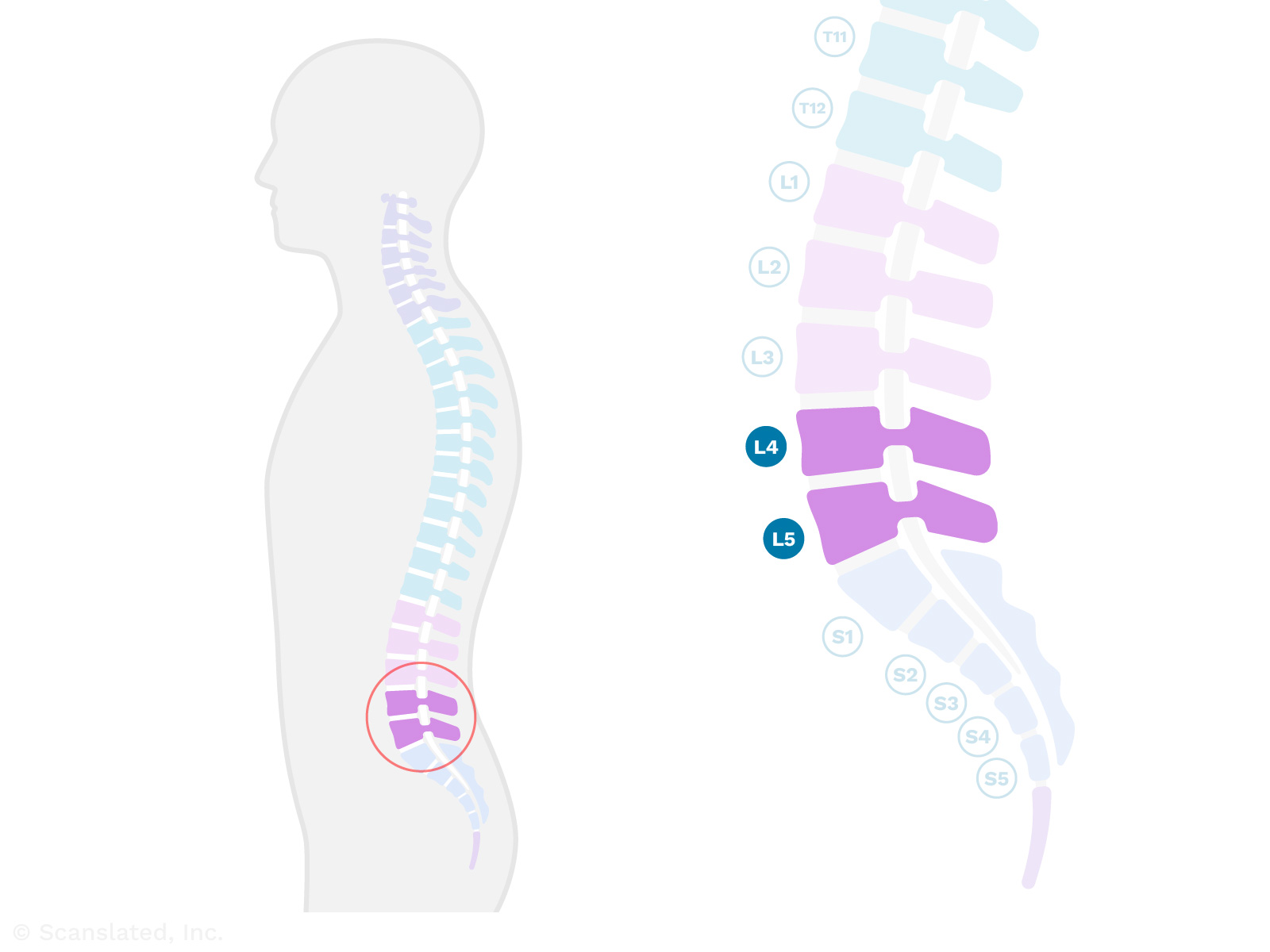
L4-L5
L4-L5
在下腰部第4和第5腰椎(脊椎骨)的位置。
腰椎通常有五块骨头, 从肋骨下方开始, 一直延伸到下背部。
L4-L5
L4-L5
Au niveau des 4ème et 5ème vertèbres lombaires (os de la colonne vertébrale) dans le bas du dos.
La colonne vertébrale lombaire, qui commence juste en dessous de la cage thoracique et se poursuit jusqu’au bas du dos, compte généralement cinq os.
Disc desiccation
When a spinal disc (soft cushion between spine bones) loses some of its water content and dries out. This happens over time with wear and tear.
disc height
The amount of space between spine bones, which is occupied by the spinal discs. This is also called “intervertebral disc space.”
Spinal discs are the soft, shock-absorbing cushions between spine bones that allow the spine to flex.
disc height
डिस्क ऊंचाई
रीढ़ की हड्डियों के बीच की जगह, जो रीढ़ की हड्डी की डिस्क द्वारा घेरी जाती है। इसे “इंटरवर्टेब्रल डिस्क स्पेस” भी कहा जाता है।
स्पाइनल डिस्क रीढ़ की हड्डियों के बीच नरम, आघात-अवशोषित कुशन होते हैं जो रीढ़ को लचीला होने देते हैं।
disc height
altura del disco
La amplitud del espacio para los discos entre los huesos de la columna vertebral (conducto vertebral intervertebral). Los discos son el material blando entre cada uno de los huesos cuadrados que forman la columna vertebral. Los discos se sitúan entre los huesos de la columna vertebral y funcionan como amortiguadores, suavizando el efecto de un impacto sobre los huesos cuando nos movemos.
disc height
椎间盘高度
椎骨之间由椎间盘占据的空间大小。这也称为”椎间盘间隙”。
椎间盘是椎骨之间的减震软垫, 使脊柱能够弯曲。
disc height
hauteur du disque
Espace entre les os de la colonne vertébrale, occupé par les disques vertébraux. Est aussi appelé espace discal intervertébral.
Les disques vertébraux sont des coussins souples qui absorbent les chocs entre les os de la colonne vertébrale et qui permettent à cette dernière de se courber.
paracentral
Slightly to the left or right of center
paracentral
पैरासेंट्रल
केंद्र से थोड़ा बायीं या दायीं ओर
paracentral
paracentral
justo a la derecha o a la izquierda de la línea media
paracentral
旁中心
中心处略微偏左或偏右
paracentral
paracentral
légèrement à gauche ou à droite du centre
disc extrusion
When the soft, gel-like material in the center of a spinal disc (cushion between spine bones) pushes out through the tough outer layer (annulus), causing a bulge.
This may not cause any problems, but in some cases can put pressure on nearby nerves and cause pain or weakness.
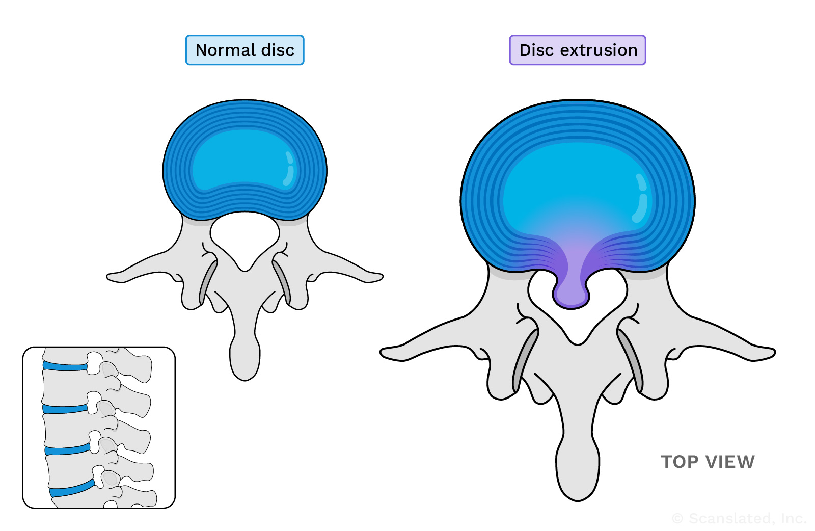
This diagram shows an example of a disc extrusion along the middle (central) part of the disc. These can also point toward the left or right.
disc extrusion
डिस्क एक्सट्रूज़न
जब रीढ़ की हड्डी की डिस्क (रीढ़ की हड्डियों के बीच कुशन) के केंद्र में स्थित नरम, जेल जैसा पदार्थ कठोर बाहरी परत (वलय) से बाहर निकलता है, तो उभार पैदा होता है।
इससे कोई समस्या तो नहीं होती, लेकिन कभी-कभी नसों पर दबाव पड़ सकता है, जिससे दर्द या कमजोरी हो सकती है।
This diagram shows an example of a disc extrusion along the middle (central) part of the disc. These can also point toward the left or right.
disc extrusion
extrusión de disco
También conocido como hernia de disco. Una extrusión (o prolapso) discal se produce cuando un desgarro en la parte exterior de un disco (material blando entre los huesos de la espalda) permite que el material gelatinoso del interior del disco se salga y provoque una protuberancia. Quizá esta protuberancia no cause ningún problema, pero en algunos casos puede ejercer presión sobre los nervios cercanos y causar dolor o debilidad.
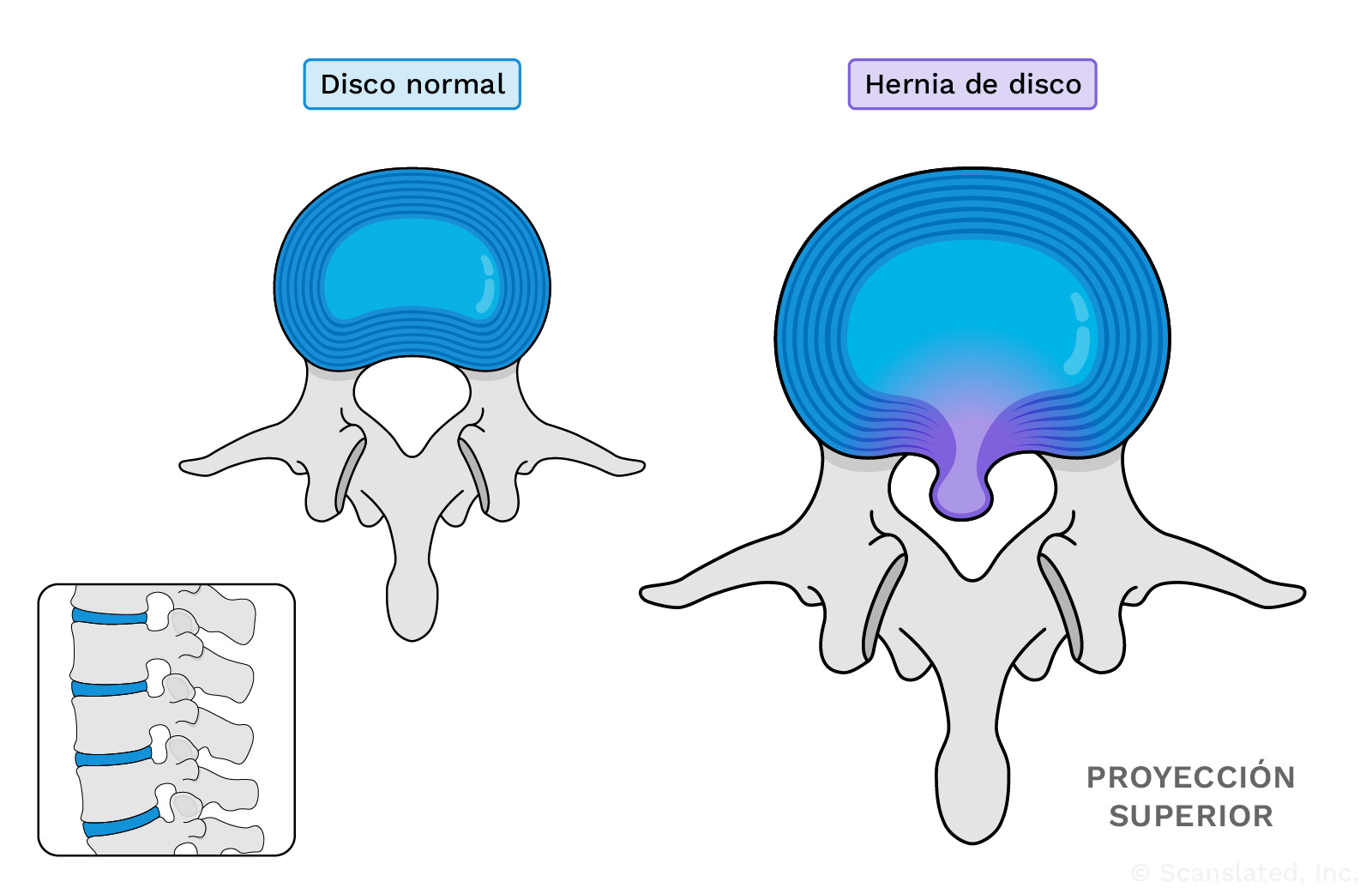
This diagram shows an example of a disc extrusion along the middle (central) part of the disc. These can also point toward the left or right.
disc extrusion
椎间盘脱出
当脊柱椎间盘(椎骨之间的缓冲垫)中心的软胶状物质从坚韧外层(纤维环)外推时, 即引起膨出。
这可能不会引起任何问题, 但有时会对附近的神经造成压迫并导致疼痛或无力。
This diagram shows an example of a disc extrusion along the middle (central) part of the disc. These can also point toward the left or right.
disc extrusion
extrusion discale
Lorsque la matière molle et gélatineuse au centre d’un disque vertébral (coussin entre les os de la colonne vertébrale) sort de la couche externe résistante (l’anneau), ce qui provoque une hernie.
Cela peut ne pas poser de problème, mais, exercer une pression sur les nerfs et provoquer des douleurs ou des faiblesses dans certains cas.
This diagram shows an example of a disc extrusion along the middle (central) part of the disc. These can also point toward the left or right.
mm
Stands for “millimeter,” which is a unit of measurement for length. A millimeter is a very small distance. For reference, there are about 25 mm in 1 inch.
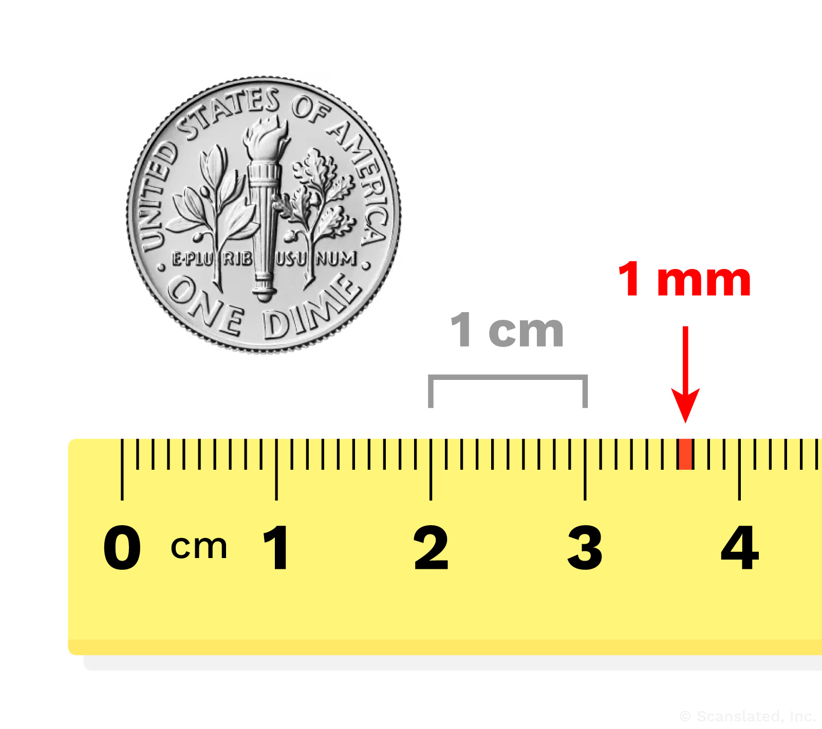
mm
mm
इसका तात्पर्य “मिलीमीटर” से है, जो लम्बाई मापने की एक इकाई है। मिलीमीटर एक बहुत छोटी दूरी है। संदर्भ के लिए, 1 इंच में लगभग 25 मिमी होते हैं।
mm
mm
abreviatura de mililitro; una unidad para medir la longitud. 1 mm es una distancia corta. Como referencia, hay unos 25 mm en 1 pulgada.
mm
毫米
代表”毫米”, 是长度的计量单位。毫米是一个非常小的距离。作为参考, 1英寸约有25毫米。
mm
mm
Il s’agit du « millimètre », qui est une unité de mesure de la longueur. Un millimètre est une très petite distance. À titre de référence, il y a environ 25 mm dans un pouce.
posteriorly
Toward the back of the body, or along the back side of something
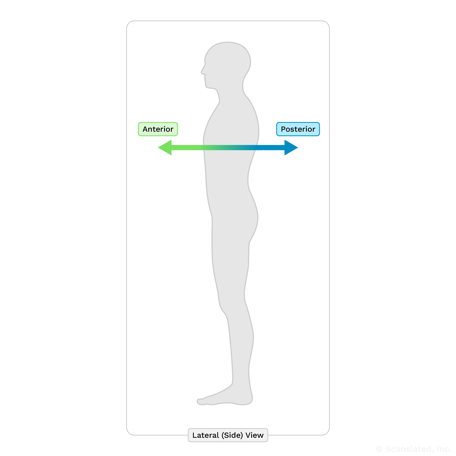
posteriorly
पीछे
शरीर के पीछे की ओर, या किसी चीज़ के पीछे की ओर
posteriorly
posterior
situado en la parte posterior o hacia la parte trasera
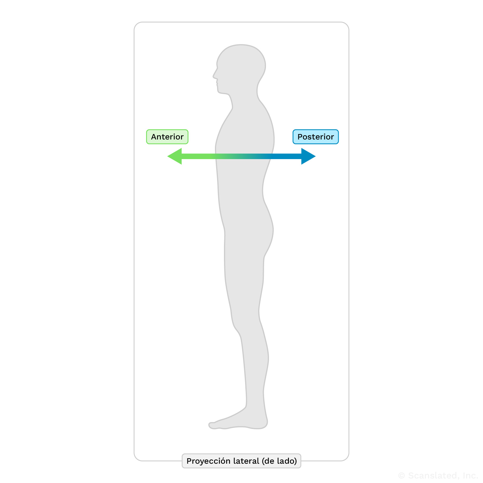
posteriorly
后部
朝向身体背侧, 或沿着某物体的背面
posteriorly
postérieurement
Vers l’arrière du corps ou le long de la face arrière de quelque chose
mm
Stands for “millimeter,” which is a unit of measurement for length. A millimeter is a very small distance. For reference, there are about 25 mm in 1 inch.
mm
mm
इसका तात्पर्य “मिलीमीटर” से है, जो लम्बाई मापने की एक इकाई है। मिलीमीटर एक बहुत छोटी दूरी है। संदर्भ के लिए, 1 इंच में लगभग 25 मिमी होते हैं।
mm
mm
abreviatura de mililitro; una unidad para medir la longitud. 1 mm es una distancia corta. Como referencia, hay unos 25 mm en 1 pulgada.
mm
毫米
代表”毫米”, 是长度的计量单位。毫米是一个非常小的距离。作为参考, 1英寸约有25毫米。
mm
mm
Il s’agit du « millimètre », qui est une unité de mesure de la longueur. Un millimètre est une très petite distance. À titre de référence, il y a environ 25 mm dans un pouce.
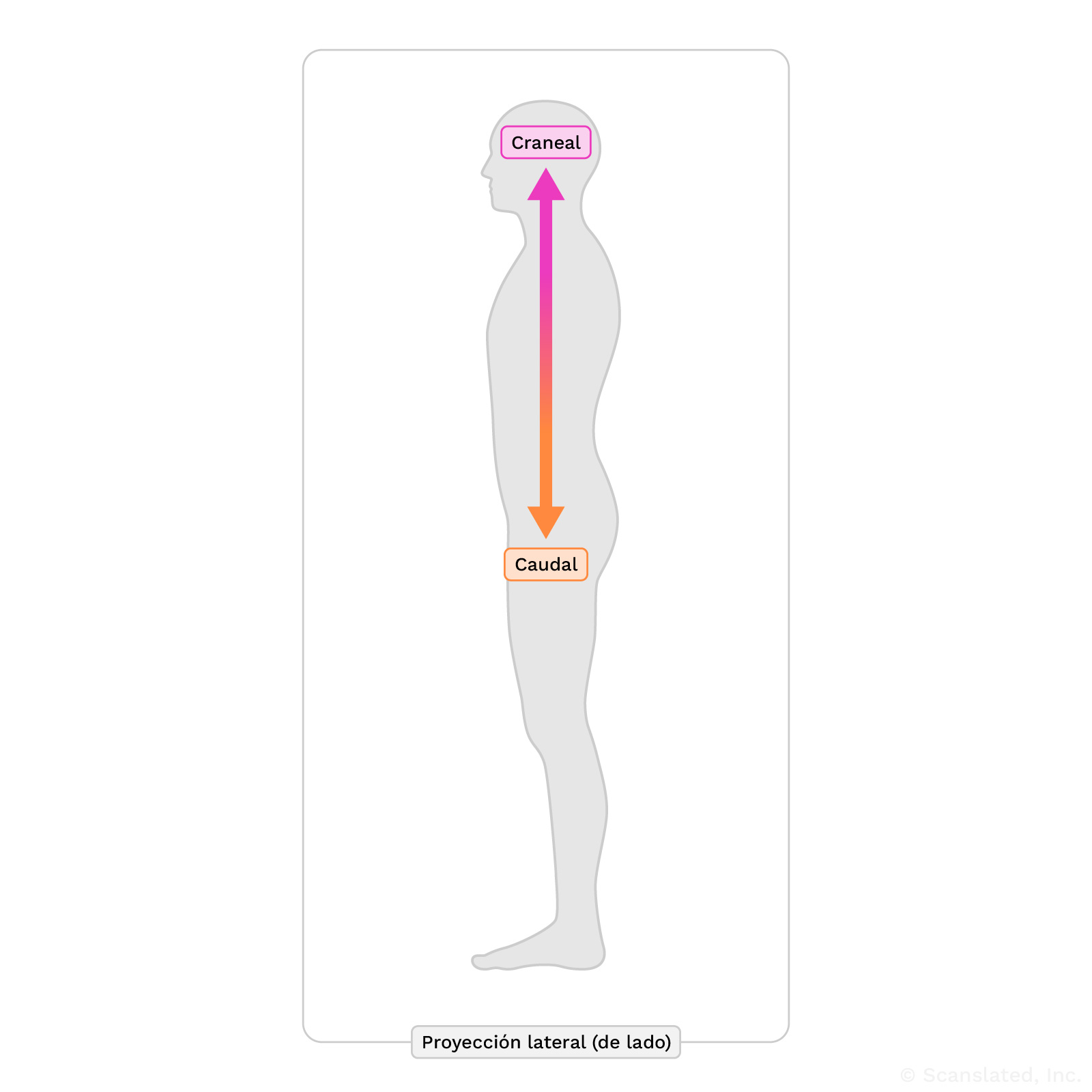
caudally
In the downward direction, toward the feet
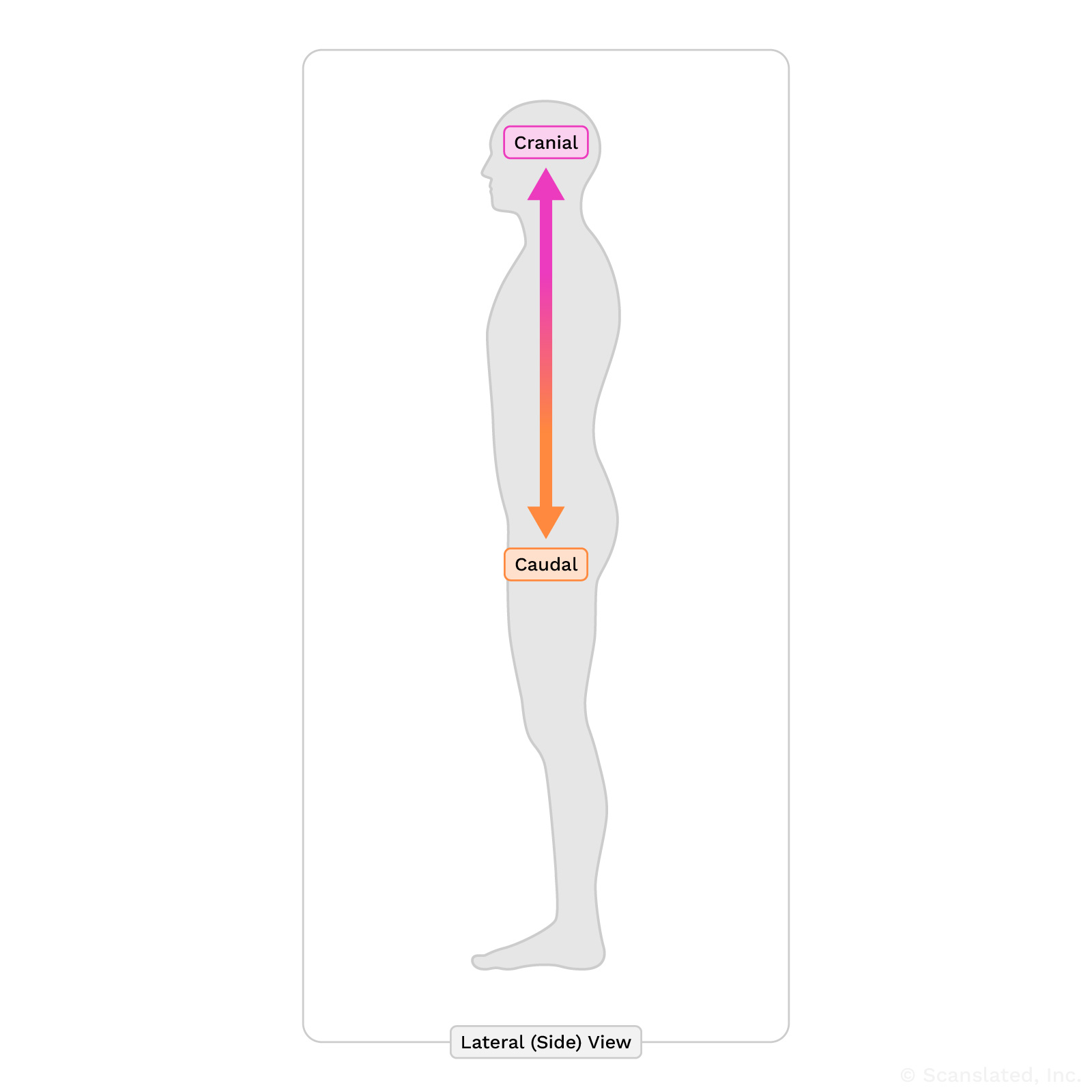
caudally
caudalmente
En la dirección hacía a los pies.
posteriorly
Toward the back of the body, or along the back side of something
posteriorly
पीछे
शरीर के पीछे की ओर, या किसी चीज़ के पीछे की ओर
posteriorly
posterior
situado en la parte posterior o hacia la parte trasera
posteriorly
后部
朝向身体背侧, 或沿着某物体的背面
posteriorly
postérieurement
Vers l’arrière du corps ou le long de la face arrière de quelque chose
nerve root
The beginning part of a nerve, just as it’s leaving the spine to go out to the body
nerve root
तंत्रिका मूल
तंत्रिका का आरंभिक भाग, जैसे ही वह रीढ़ की हड्डी को छोड़कर शरीर में जाती है
nerve root
raíz nerviosa
La parte inicial de un nervio, justo cuando está saliendo de la columna vertebral para salir al cuerpo.
nerve root
神经根
神经的起始段, 即刚从脊柱伸出的一段, 其后延伸至身体各部位
nerve root
racine nerveuse
Partie initiale d’un nerf, au moment où il quitte la colonne vertébrale pour se diriger vers le corps.
lateral
Toward one side (left or right)
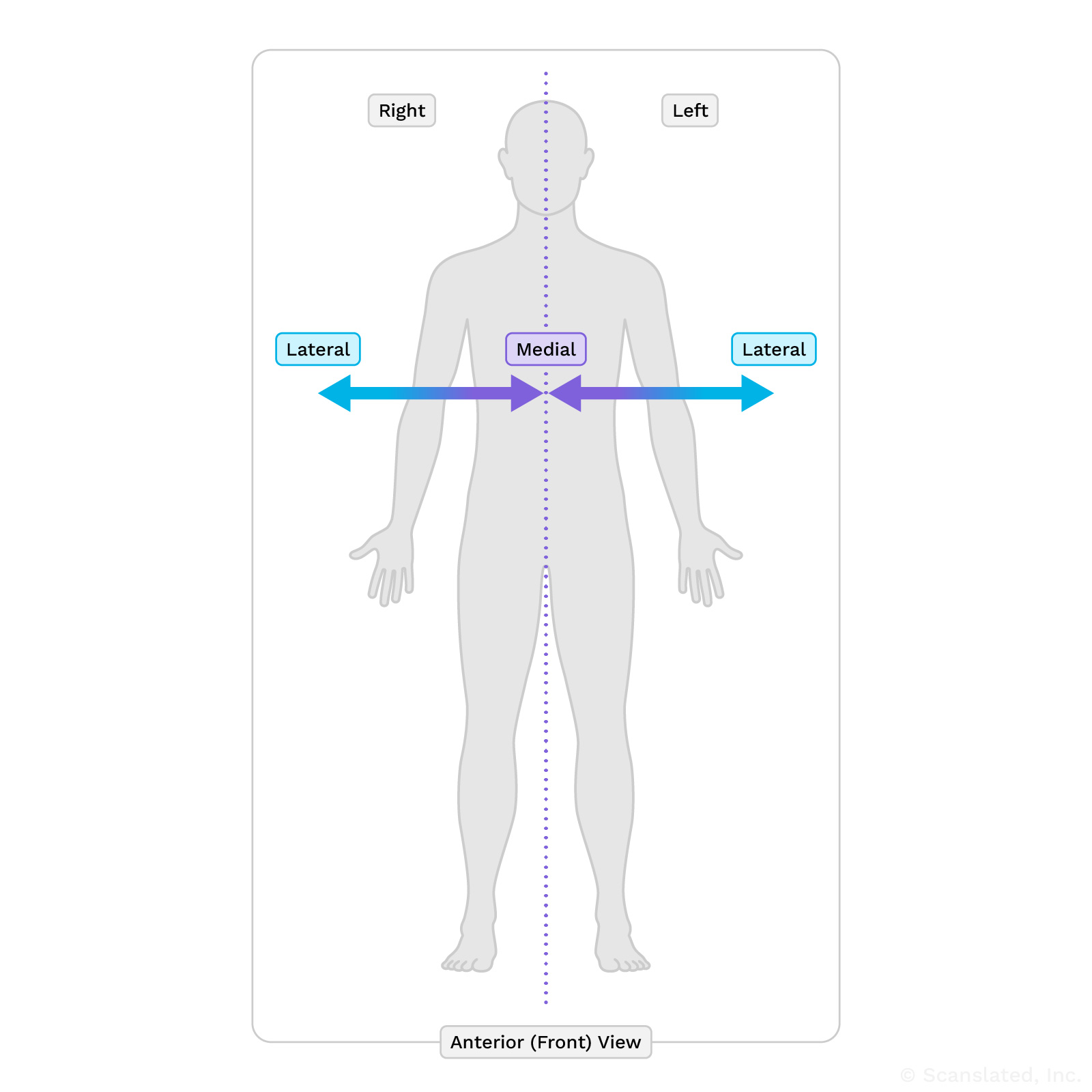
lateral
पार्श्व
एक ओर (बाएं या दाएं)
lateral
lateral o externo
hacia un lado (izquierda o derecha)
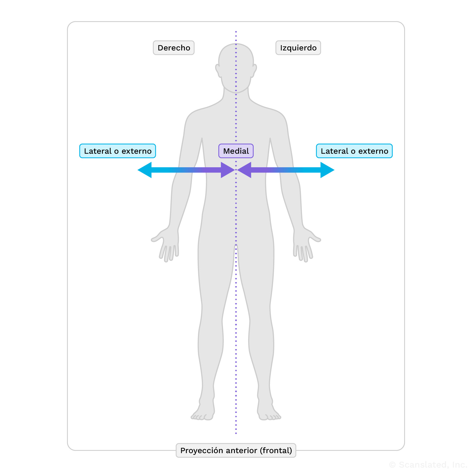
lateral
侧边的
偏向一侧(左侧或右侧)
lateral
latéral(e)
Vers un côté (gauche ou droit)
recess
A space in the body, often between or around organs
recess
अवकाश
शरीर में एक स्थान, जो प्रायः अंगों के बीच या उनके आस-पास होता है
recess
hueco
otro término para designar un espacio en el cuerpo, a menudo entre o alrededor de otros órganos.
recess
隐窝
人体中通常位于器官之间或周围的一个空腔
recess
récessus
Espace dans le corps, souvent entre ou autour des organes.
Moderate
Not mild, but not severe; in the middle
bilateral facet arthropathy
Wear-and-tear changes of the facet joints, which are the small joints that link the vertebrae (spine bones) together. The facet joints allow the spine to bend forward and backward.
These changes are common with age, and can sometimes be a cause for back or neck pain.
Facet joints are highlighted here in blue. Facet joints are present all along the spine; the lumbar section is shown here as an example.
bilateral facet arthropathy
द्विपक्षीय पहलू आर्थ्रोपैथी
पहलू जोड़ों में टूट-फूट से होने वाले परिवर्तन, जो कि छोटे जोड़ होते हैं जो कशेरुकाओं (रीढ़ की हड्डियों) को एक साथ जोड़ते हैं। पहलू जोड़ रीढ़ को आगे और पीछे झुकने की अनुमति देते हैं।
ये परिवर्तन उम्र के साथ आम हैं और कभी-कभी पीठ या गर्दन में दर्द का कारण भी बन सकते हैं।
Facet joints are highlighted here in blue. Facet joints are present all along the spine; the lumbar section is shown here as an example.
bilateral facet arthropathy
artropatía facetaria bilateral
Cambios por desgaste de las articulaciones pequeñas (facetarias) que unen a los huesos de la columna vertebral (vertebras). Estos cambios son habituales con el envejecimiento. A veces puede ser una causa de dolor de espalda.
Facet joints are highlighted here in blue. Facet joints are present all along the spine; the lumbar section is shown here as an example.
bilateral facet arthropathy
双侧小关节病
小关节(连接脊柱骨 [椎骨] 的小关节)的磨损性改变。小关节使脊柱可以向前和向后弯曲。
随着年龄的增长, 这种改变很常见, 有时会引起背部或颈部疼痛。
Facet joints are highlighted here in blue. Facet joints are present all along the spine; the lumbar section is shown here as an example.
bilateral facet arthropathy
arthropathie facettaire bilatérale
Usure des facettes vertébrales qui sont de petites articulations reliant les vertèbres (les os de la colonne vertébrale) ensemble. Les facettes vertébrales permettent à la colonne vertébrale de se courber en avant et en arrière.
Ces modifications sont courantes avec l’âge et peuvent parfois être la cause de douleurs dans le dos ou dans le cou.
Facet joints are highlighted here in blue. Facet joints are present all along the spine; the lumbar section is shown here as an example.
ligamentum flavum
A ligament (strong fibrous band) that runs along the back side of the vertebrae (spine bones). It stabilizes the spine while allowing it to bend.
Sometimes, the ligamentum flavum can become thickened and push into the spinal canal (space where the nerves travel), which can cause pain, numbness, or weakness. This happens due to wear and tear on the spine over time.
ligamentum flavum
लिगामेंटम फ्लेवम
एक लिगामेंट (मजबूत रेशेदार बैंड) जो कशेरुकाओं (रीढ़ की हड्डियों) के पीछे की ओर चलता है। यह रीढ़ की हड्डी को स्थिर रखता है तथा उसे झुकने की अनुमति देता है।
कभी-कभी, लिगामेंटम फ्लेवम मोटा हो सकता है और स्पाइनल कैनाल (वह स्थान जहां तंत्रिकाएं यात्रा करती हैं) में धकेल सकता है, जिससे दर्द, सुन्नता या कमजोरी हो सकती है। ऐसा समय के साथ रीढ़ की हड्डी में होने वाली टूट-फूट के कारण होता है।
ligamentum flavum
ligamento amarillo
El ligamento amarillo es un ligamento fibroso y resistente o una banda a lo largo de la parte posterior de los huesos de la columna vertebral, que mantiene los huesos unidos y alineados entre sí.
ligamentum flavum
黄韧带
沿着脊柱骨(椎骨)背面移行的韧带(强韧的纤维带)。它既能稳定脊柱, 又能让脊柱弯曲。
有时, 黄韧带会变厚并挤入椎管(神经移行的空间), 从而导致疼痛、麻木或无力。这种情况的出现是由于脊柱逐渐磨损造成的。
ligamentum flavum
ligament jaune
Ligament (une bande fibreuse résistante) qui longe la face postérieure des vertèbres (os de la colonne vertébrale). Il stabilise la colonne vertébrale tout en lui permettant de se courber.
Parfois le ligament jaune épaissit et pousse dans le canal rachidien (l’espace où courent les nerfs), ce qui peut cause de la douleur, des engourdissements ou de la faiblesse. Cela survient avec l’usure de la colonne vertébrale avec le temps.
thickening
Widening; wider than usual
thickening
और अधिक मोटा होना
चौड़ा होना; सामान्य से अधिक चौड़ा होना
thickening
espesamiento
ampliación, más amplio de lo habitual
thickening
增厚
变宽; 比正常宽
thickening
épaississement
élargissement ; plus large que la normale
spinal canal stenosis
Narrowing of the spinal canal (the space where the spinal cord and nerves travel through the spine bones). This can put pressure on the spinal cord and nerves, which can lead to symptoms like pain, numbness, or weakness.
Stenosis (narrowing) can be caused by a bulging disc, bone spurs, or enlargement of the ligaments in the back of the spine.
neural foraminal narrowing
Narrowing (stenosis) of the neural foramina, which can be caused by bone spurs or bulging discs.
A “foramen” (plural: “foramina”) is a normal hole or tunnel in a bone.
In the spine, neural foramina are openings on either side of the vertebrae (spine bones) where the nerves travel. Nerves exit the spinal canal through these openings at each level of the spine, going out to the left and right sides of the body.
Narrowing of these openings can sometimes be a cause for pain.
neural foraminal narrowing
तंत्रिका छिद्र का संकुचन
तंत्रिका छिद्रों का संकुचित होना (स्टेनोसिस), जो अस्थि स्पर्स या उभरी हुई डिस्क के कारण हो सकता है।
एक “फोरैमन” (बहुवचन: “फोरैमिना”) हड्डी में एक सामान्य छेद या सुरंग है।
रीढ़ में, न्यूरल फोरैमिना कशेरुकाओं (रीढ़ की हड्डियों) के दोनों ओर खुले होते हैं जहां तंत्रिकाएं यात्रा करती हैं। रीढ़ की हड्डी के प्रत्येक स्तर पर इन छिद्रों के माध्यम से नसें रीढ़ की हड्डी की नहर से बाहर निकलती हैं, शरीर के बाएँ और दाएँ तरफ जाती हैं।
इन छिद्रों का संकुचित होना कभी-कभी दर्द का कारण बन सकता है।
neural foraminal narrowing
pinzamiento del agujero intervertebral
Estrechez de las aberturas en cada lado de los huesos de la columna vertebral por donde recorren los nervios. Los nervios salen del canal espinal por cada nivel de la columna vertebral y transmiten señales de movimiento y de sensación a las diferentes partes del cuerpo. A cada nivel, hay un nervio que se dirige al lado derecho, y un nervio que se dirige al lado izquierdo del cuerpo. Estos nervios recorren por el agujero intervertebral en cada lado al salir de la columna vertebral. El estrechamiento de estas aberturas puede ser causado por espolones óseos o hernias discales.
neural foraminal narrowing
神经孔狭窄
神经孔变窄(狭窄), 可能由骨刺或椎间盘膨出引起。
骨骼中存在正常的孔洞或隧道, 称之为”孔”。
在脊柱中, 神经孔是脊柱骨(椎骨)一侧的有神经穿过的开口。神经通过每个脊柱水平的这些开口离开椎管, 通向身体的左右两侧。
这些开口变窄有时会导致疼痛。
neural foraminal narrowing
rétrécissement du foramen neural
Le rétrécissement (sténose) du foramen neural peut être causé par des épines osseuses ou des hernies discales.
Le « foramen » (pluriel : « foramina » ) est un trou ou un tunnel normal dans un os.
Dans la colonne vertébrale, les foramina neuraux sont des ouvertures de chaque côté des vertèbres (les os de la colonne vertébrale) où passent les nerfs. Les nerfs sortent du canal rachidien par ces ouvertures à chaque niveau de la colonne vertébrale et partent à droite et à gauche du corps.
Le rétrécissement de ces ouvertures peut parfois provoquer de la douleur.
neural foraminal narrowing
Narrowing (stenosis) of the neural foramina, which can be caused by bone spurs or bulging discs.
A “foramen” (plural: “foramina”) is a normal hole or tunnel in a bone.
In the spine, neural foramina are openings on either side of the vertebrae (spine bones) where the nerves travel. Nerves exit the spinal canal through these openings at each level of the spine, going out to the left and right sides of the body.
Narrowing of these openings can sometimes be a cause for pain.
neural foraminal narrowing
तंत्रिका छिद्र का संकुचन
तंत्रिका छिद्रों का संकुचित होना (स्टेनोसिस), जो अस्थि स्पर्स या उभरी हुई डिस्क के कारण हो सकता है।
एक “फोरैमन” (बहुवचन: “फोरैमिना”) हड्डी में एक सामान्य छेद या सुरंग है।
रीढ़ में, न्यूरल फोरैमिना कशेरुकाओं (रीढ़ की हड्डियों) के दोनों ओर खुले होते हैं जहां तंत्रिकाएं यात्रा करती हैं। रीढ़ की हड्डी के प्रत्येक स्तर पर इन छिद्रों के माध्यम से नसें रीढ़ की हड्डी की नहर से बाहर निकलती हैं, शरीर के बाएँ और दाएँ तरफ जाती हैं।
इन छिद्रों का संकुचित होना कभी-कभी दर्द का कारण बन सकता है।
neural foraminal narrowing
pinzamiento del agujero intervertebral
Estrechez de las aberturas en cada lado de los huesos de la columna vertebral por donde recorren los nervios. Los nervios salen del canal espinal por cada nivel de la columna vertebral y transmiten señales de movimiento y de sensación a las diferentes partes del cuerpo. A cada nivel, hay un nervio que se dirige al lado derecho, y un nervio que se dirige al lado izquierdo del cuerpo. Estos nervios recorren por el agujero intervertebral en cada lado al salir de la columna vertebral. El estrechamiento de estas aberturas puede ser causado por espolones óseos o hernias discales.
neural foraminal narrowing
神经孔狭窄
神经孔变窄(狭窄), 可能由骨刺或椎间盘膨出引起。
骨骼中存在正常的孔洞或隧道, 称之为”孔”。
在脊柱中, 神经孔是脊柱骨(椎骨)一侧的有神经穿过的开口。神经通过每个脊柱水平的这些开口离开椎管, 通向身体的左右两侧。
这些开口变窄有时会导致疼痛。
neural foraminal narrowing
rétrécissement du foramen neural
Le rétrécissement (sténose) du foramen neural peut être causé par des épines osseuses ou des hernies discales.
Le « foramen » (pluriel : « foramina » ) est un trou ou un tunnel normal dans un os.
Dans la colonne vertébrale, les foramina neuraux sont des ouvertures de chaque côté des vertèbres (les os de la colonne vertébrale) où passent les nerfs. Les nerfs sortent du canal rachidien par ces ouvertures à chaque niveau de la colonne vertébrale et partent à droite et à gauche du corps.
Le rétrécissement de ces ouvertures peut parfois provoquer de la douleur.
L5-S1
Where the last lumbar vertebra (L5) meets the top of the sacrum (S1).
The sacrum is the large, triangle-shaped “sitting bone” that makes up the back part of the pelvis and is connected to the lower (lumbar) spine. It’s made up of five bones, which usually fuse (join together) during early adulthood.
L5-S1
L5-S1
जहां अंतिम कटि कशेरुका (L5) त्रिकास्थि (S1) के शीर्ष से मिलती है।
त्रिकास्थि बड़ी, त्रिकोण के आकार की “बैठने वाली हड्डी” है जो श्रोणि के पीछे का हिस्सा बनाती है और निचली (काठ) रीढ़ से जुड़ी होती है। यह पाँच हड्डियों से बनी होती है, जो आमतौर पर प्रारंभिक वयस्कता के दौरान जुड़ जाती हैं (एक साथ जुड़ जाती हैं)।
L5-S1
L5-S1
El espacio entre el último hueso lumbar (L5) y el primer hueso sacro (S1). Hay un disco (cojín amortiguador blando) entre los dos huesos y las articulaciones a lo largo de la parte posterior que une los huesos. El nivel L5-S1 se encuentra en la parte más baja de la espalda, justo por encima de los huesos del asiento.
L5-S1
L5-S1
最后一节腰椎(L5)与骶骨顶部(S1)的交接处。
骶骨是一块三角形的大”坐骨”, 构成骨盆的后部, 与下部(腰椎)脊柱相连。它由五块骨头组成, 通常在成年早期会融合(连接)在一起。
L5-S1
L5-S1
Point de rencontre entre la dernière vertèbre lombaire (L5) et la partie supérieure du sacrum (S1).
Le sacrum est le grand « os de l’assise » en forme de triangle qui constitue la partie arrière du bassin et est relié à la colonne vertébrale inférieure (lombaire). Il est composé de cinq os, qui fusionnent généralement au début de l’âge adulte.
Disc desiccation
When a spinal disc (soft cushion between spine bones) loses some of its water content and dries out. This happens over time with wear and tear.
moderate
Not mild, but not severe; in the middle
disc height
The amount of space between spine bones, which is occupied by the spinal discs. This is also called “intervertebral disc space.”
Spinal discs are the soft, shock-absorbing cushions between spine bones that allow the spine to flex.
disc height
डिस्क ऊंचाई
रीढ़ की हड्डियों के बीच की जगह, जो रीढ़ की हड्डी की डिस्क द्वारा घेरी जाती है। इसे “इंटरवर्टेब्रल डिस्क स्पेस” भी कहा जाता है।
स्पाइनल डिस्क रीढ़ की हड्डियों के बीच नरम, आघात-अवशोषित कुशन होते हैं जो रीढ़ को लचीला होने देते हैं।
disc height
altura del disco
La amplitud del espacio para los discos entre los huesos de la columna vertebral (conducto vertebral intervertebral). Los discos son el material blando entre cada uno de los huesos cuadrados que forman la columna vertebral. Los discos se sitúan entre los huesos de la columna vertebral y funcionan como amortiguadores, suavizando el efecto de un impacto sobre los huesos cuando nos movemos.
disc height
椎间盘高度
椎骨之间由椎间盘占据的空间大小。这也称为”椎间盘间隙”。
椎间盘是椎骨之间的减震软垫, 使脊柱能够弯曲。
disc height
hauteur du disque
Espace entre les os de la colonne vertébrale, occupé par les disques vertébraux. Est aussi appelé espace discal intervertébral.
Les disques vertébraux sont des coussins souples qui absorbent les chocs entre les os de la colonne vertébrale et qui permettent à cette dernière de se courber.
Broad-based disc bulge
A disc bulge is an outpouching of the spinal disc (the soft cushion between spine bones). “Broad-based” generally means involving more than 25% of the outer contour of the disc.
Disc bulges are fairly common, and may not cause any problems, but can sometimes put pressure on nerves, causing pain or weakness.
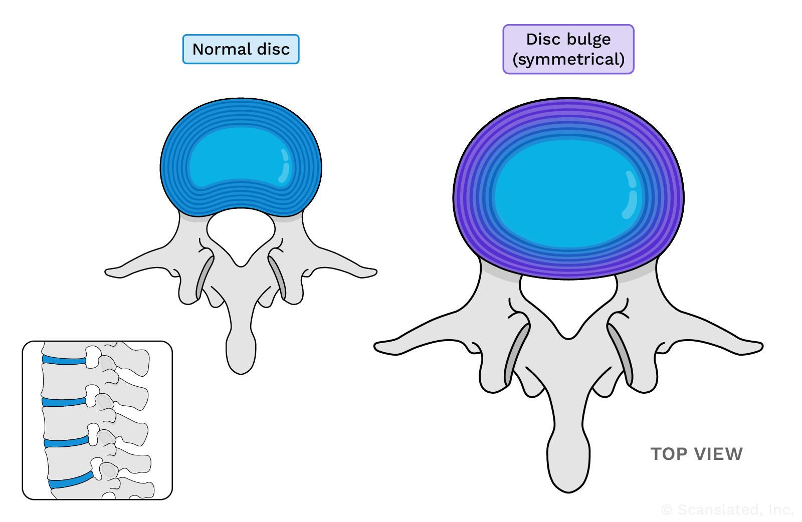
focal
Seen in a specific spot or location; something you can point to
focal
केंद्रीय
किसी विशिष्ट स्थान या जगह पर देखा गया; ऐसी चीज़ जिसकी ओर आप इशारा कर सकें
focal
focal
visto en un punto o lugar específico, algo que se pueda indicar
focal
局灶性
在特定点或位置看到的; 可以指向的东西
focal
focal
Vu dans un endroit ou un lieu spécifique ; quelque chose que l’on peut montrer du doigt
bilateral facet arthropathy
Wear-and-tear changes of the facet joints, which are the small joints that link the vertebrae (spine bones) together. The facet joints allow the spine to bend forward and backward.
These changes are common with age, and can sometimes be a cause for back or neck pain.
Facet joints are highlighted here in blue. Facet joints are present all along the spine; the lumbar section is shown here as an example.
bilateral facet arthropathy
द्विपक्षीय पहलू आर्थ्रोपैथी
पहलू जोड़ों में टूट-फूट से होने वाले परिवर्तन, जो कि छोटे जोड़ होते हैं जो कशेरुकाओं (रीढ़ की हड्डियों) को एक साथ जोड़ते हैं। पहलू जोड़ रीढ़ को आगे और पीछे झुकने की अनुमति देते हैं।
ये परिवर्तन उम्र के साथ आम हैं और कभी-कभी पीठ या गर्दन में दर्द का कारण भी बन सकते हैं।
Facet joints are highlighted here in blue. Facet joints are present all along the spine; the lumbar section is shown here as an example.
bilateral facet arthropathy
artropatía facetaria bilateral
Cambios por desgaste de las articulaciones pequeñas (facetarias) que unen a los huesos de la columna vertebral (vertebras). Estos cambios son habituales con el envejecimiento. A veces puede ser una causa de dolor de espalda.
Facet joints are highlighted here in blue. Facet joints are present all along the spine; the lumbar section is shown here as an example.
bilateral facet arthropathy
双侧小关节病
小关节(连接脊柱骨 [椎骨] 的小关节)的磨损性改变。小关节使脊柱可以向前和向后弯曲。
随着年龄的增长, 这种改变很常见, 有时会引起背部或颈部疼痛。
Facet joints are highlighted here in blue. Facet joints are present all along the spine; the lumbar section is shown here as an example.
bilateral facet arthropathy
arthropathie facettaire bilatérale
Usure des facettes vertébrales qui sont de petites articulations reliant les vertèbres (les os de la colonne vertébrale) ensemble. Les facettes vertébrales permettent à la colonne vertébrale de se courber en avant et en arrière.
Ces modifications sont courantes avec l’âge et peuvent parfois être la cause de douleurs dans le dos ou dans le cou.
Facet joints are highlighted here in blue. Facet joints are present all along the spine; the lumbar section is shown here as an example.
spinal canal stenosis
Narrowing of the spinal canal (the space where the spinal cord and nerves travel through the spine bones). This can put pressure on the spinal cord and nerves, which can lead to symptoms like pain, numbness, or weakness.
Stenosis (narrowing) can be caused by a bulging disc, bone spurs, or enlargement of the ligaments in the back of the spine.
bilateral
Involving or referring to both the left and right sides
neural foraminal narrowing
Narrowing (stenosis) of the neural foramina, which can be caused by bone spurs or bulging discs.
A “foramen” (plural: “foramina”) is a normal hole or tunnel in a bone.
In the spine, neural foramina are openings on either side of the vertebrae (spine bones) where the nerves travel. Nerves exit the spinal canal through these openings at each level of the spine, going out to the left and right sides of the body.
Narrowing of these openings can sometimes be a cause for pain.
neural foraminal narrowing
तंत्रिका छिद्र का संकुचन
तंत्रिका छिद्रों का संकुचित होना (स्टेनोसिस), जो अस्थि स्पर्स या उभरी हुई डिस्क के कारण हो सकता है।
एक “फोरैमन” (बहुवचन: “फोरैमिना”) हड्डी में एक सामान्य छेद या सुरंग है।
रीढ़ में, न्यूरल फोरैमिना कशेरुकाओं (रीढ़ की हड्डियों) के दोनों ओर खुले होते हैं जहां तंत्रिकाएं यात्रा करती हैं। रीढ़ की हड्डी के प्रत्येक स्तर पर इन छिद्रों के माध्यम से नसें रीढ़ की हड्डी की नहर से बाहर निकलती हैं, शरीर के बाएँ और दाएँ तरफ जाती हैं।
इन छिद्रों का संकुचित होना कभी-कभी दर्द का कारण बन सकता है।
neural foraminal narrowing
pinzamiento del agujero intervertebral
Estrechez de las aberturas en cada lado de los huesos de la columna vertebral por donde recorren los nervios. Los nervios salen del canal espinal por cada nivel de la columna vertebral y transmiten señales de movimiento y de sensación a las diferentes partes del cuerpo. A cada nivel, hay un nervio que se dirige al lado derecho, y un nervio que se dirige al lado izquierdo del cuerpo. Estos nervios recorren por el agujero intervertebral en cada lado al salir de la columna vertebral. El estrechamiento de estas aberturas puede ser causado por espolones óseos o hernias discales.
neural foraminal narrowing
神经孔狭窄
神经孔变窄(狭窄), 可能由骨刺或椎间盘膨出引起。
骨骼中存在正常的孔洞或隧道, 称之为”孔”。
在脊柱中, 神经孔是脊柱骨(椎骨)一侧的有神经穿过的开口。神经通过每个脊柱水平的这些开口离开椎管, 通向身体的左右两侧。
这些开口变窄有时会导致疼痛。
neural foraminal narrowing
rétrécissement du foramen neural
Le rétrécissement (sténose) du foramen neural peut être causé par des épines osseuses ou des hernies discales.
Le « foramen » (pluriel : « foramina » ) est un trou ou un tunnel normal dans un os.
Dans la colonne vertébrale, les foramina neuraux sont des ouvertures de chaque côté des vertèbres (les os de la colonne vertébrale) où passent les nerfs. Les nerfs sortent du canal rachidien par ces ouvertures à chaque niveau de la colonne vertébrale et partent à droite et à gauche du corps.
Le rétrécissement de ces ouvertures peut parfois provoquer de la douleur.
impingement
Abnormal pinching or squeezing
impingement
अतिक्रमण
असामान्य चुभन या दबाव
impingement
pinzamiento o conflicto de espacio
Pellizcos o apretones anormales
impingement
撞击
异常的捏夹或挤压
impingement
empiètement
Pincement ou compression anormale
nerve roots
The “nerve root” is the beginning part of a nerve, just as it’s leaving the spine to go out to the body.
nerve roots
तंत्रिका जड़ें
तंत्रिका मूल तंत्रिका का प्रारंभिक भाग है, जो रीढ़ की हड्डी को छोड़कर शरीर में जाती है।
nerve roots
raíces nerviosas
Una raíz nerviosa es la parte inicial de un nervio, justo cuando está saliendo de la columna vertebral para salir al cuerpo.
nerve roots
神经根
“神经根”是神经的起始段, 即刚从脊柱伸出的一段, 其后延伸至身体各部位。
nerve roots
racines nerveuses
La« racine nerveuse » est la partie initiale d’un nerf, au moment où il quitte la colonne vertébrale pour se diriger vers le corps.
Paraspinal
Next to or around the spine. There are muscles, fat, and other soft tissues in this area.
Soft Tissues
Body tissues other than bone. Soft tissues include muscle, fat, and skin (among others).
Soft Tissues
नरम ऊतक
हड्डी के अलावा अन्य शरीर के ऊतक। नरम ऊतकों में मांसपेशियां, वसा और त्वचा (अन्य) शामिल हैं।
Soft Tissues
partes blandas
Tejidos corporales (del cuerpo) distinto del hueso. Las partes blandas incluyen el músculo, la grasa y la piel (entre otros).
Soft Tissues
软组织
除骨骼以外的身体组织。软组织包括肌肉、脂肪和皮肤等。
Soft Tissues
tissus mous
Tissus du corps autre que les os. Les tissus mous comprennent les muscles, le tissu adipeux et la peau (entre autres).
Unremarkable
Normal; imaging looks as expected
IMPRESSION
The Impression is a concise summary of what was seen on the images, containing the most important or relevant observations.
IMPRESSION
छाप
रेडियोलॉजिस्ट ने स्कैन में जो देखा उसका सारांश, जिसमें सबसे महत्वपूर्ण या प्रासंगिक अवलोकन शामिल हैं।
रिपोर्ट में जो कुछ भी टिप्पणी की गई थी, उसे इंप्रेशन में शामिल नहीं किया जाएगा - केवल सबसे महत्वपूर्ण चीजें जो रेडियोलॉजिस्ट संचार करना चाहता है।
IMPRESSION
impresión diagnóstica
La impresión es el resumen de lo que el radiólogo ha observado en la exploración, y contiene las observaciones más importantes o relevantes. No todo lo que el radiólogo comentó se incluirá en la impresión, sólo lo más importante que el radiólogo quiere comunicar.
IMPRESSION
影像学所见总结
放射科医生对扫描结果的总结, 包含最重要或最相关的观察结果。
并非报告中评论的所有内容都会包含在影像学所见总结中, 只有放射科医生希望传达的最重要的内容才会包含在其中。
IMPRESSION
impression
Résumé de ce que le radiologue a vu sur le scanner, contenant les observations les plus importantes ou les plus pertinentes.
Tout ce qui a été commenté dans le rapport ne sera pas inclus dans l’impression - seuls les éléments les plus importants que le radiologue souhaite communiquer.
L4-L5
At the level of the 4th and 5th lumbar vertebrae (spine bones) in the lower back.
There are usually five bones in the lumbar spine, which begins just below the rib cage and continues down through the lower back.
L4-L5
L4-L5
निचली पीठ में चौथी और पांचवीं काठ कशेरुका (रीढ़ की हड्डी) के स्तर पर।
काठ की रीढ़ में आमतौर पर पांच हड्डियां होती हैं, जो पसलियों के पिंजरे के ठीक नीचे से शुरू होती हैं और पीठ के निचले हिस्से से नीचे तक जाती हैं।
L4-L5
L4-L5
Entre los cuerpos vertebrales lumbares 4º y 5º (huesos de la parte baja de la espalda). Hay un disco (cojín amortiguador blando) entre los dos huesos y las articulaciones a lo largo de la parte posterior que une los huesos.
L4-L5
L4-L5
在下腰部第4和第5腰椎(脊椎骨)的位置。
腰椎通常有五块骨头, 从肋骨下方开始, 一直延伸到下背部。
L4-L5
L4-L5
Au niveau des 4ème et 5ème vertèbres lombaires (os de la colonne vertébrale) dans le bas du dos.
La colonne vertébrale lombaire, qui commence juste en dessous de la cage thoracique et se poursuit jusqu’au bas du dos, compte généralement cinq os.
paracentral
Slightly to the left or right of center
paracentral
पैरासेंट्रल
केंद्र से थोड़ा बायीं या दायीं ओर
paracentral
paracentral
justo a la derecha o a la izquierda de la línea media
paracentral
旁中心
中心处略微偏左或偏右
paracentral
paracentral
légèrement à gauche ou à droite du centre
disc extrusion
When the soft, gel-like material in the center of a spinal disc (cushion between spine bones) pushes out through the tough outer layer (annulus), causing a bulge.
This may not cause any problems, but in some cases can put pressure on nearby nerves and cause pain or weakness.
This diagram shows an example of a disc extrusion along the middle (central) part of the disc. These can also point toward the left or right.
disc extrusion
डिस्क एक्सट्रूज़न
जब रीढ़ की हड्डी की डिस्क (रीढ़ की हड्डियों के बीच कुशन) के केंद्र में स्थित नरम, जेल जैसा पदार्थ कठोर बाहरी परत (वलय) से बाहर निकलता है, तो उभार पैदा होता है।
इससे कोई समस्या तो नहीं होती, लेकिन कभी-कभी नसों पर दबाव पड़ सकता है, जिससे दर्द या कमजोरी हो सकती है।
This diagram shows an example of a disc extrusion along the middle (central) part of the disc. These can also point toward the left or right.
disc extrusion
extrusión de disco
También conocido como hernia de disco. Una extrusión (o prolapso) discal se produce cuando un desgarro en la parte exterior de un disco (material blando entre los huesos de la espalda) permite que el material gelatinoso del interior del disco se salga y provoque una protuberancia. Quizá esta protuberancia no cause ningún problema, pero en algunos casos puede ejercer presión sobre los nervios cercanos y causar dolor o debilidad.
This diagram shows an example of a disc extrusion along the middle (central) part of the disc. These can also point toward the left or right.
disc extrusion
椎间盘脱出
当脊柱椎间盘(椎骨之间的缓冲垫)中心的软胶状物质从坚韧外层(纤维环)外推时, 即引起膨出。
这可能不会引起任何问题, 但有时会对附近的神经造成压迫并导致疼痛或无力。
This diagram shows an example of a disc extrusion along the middle (central) part of the disc. These can also point toward the left or right.
disc extrusion
extrusion discale
Lorsque la matière molle et gélatineuse au centre d’un disque vertébral (coussin entre les os de la colonne vertébrale) sort de la couche externe résistante (l’anneau), ce qui provoque une hernie.
Cela peut ne pas poser de problème, mais, exercer une pression sur les nerfs et provoquer des douleurs ou des faiblesses dans certains cas.
This diagram shows an example of a disc extrusion along the middle (central) part of the disc. These can also point toward the left or right.
nerve root
The beginning part of a nerve, just as it’s leaving the spine to go out to the body
nerve root
तंत्रिका मूल
तंत्रिका का आरंभिक भाग, जैसे ही वह रीढ़ की हड्डी को छोड़कर शरीर में जाती है
nerve root
raíz nerviosa
La parte inicial de un nervio, justo cuando está saliendo de la columna vertebral para salir al cuerpo.
nerve root
神经根
神经的起始段, 即刚从脊柱伸出的一段, 其后延伸至身体各部位
nerve root
racine nerveuse
Partie initiale d’un nerf, au moment où il quitte la colonne vertébrale pour se diriger vers le corps.
leg
In common usage, “leg” refers to the entire lower limb (from the thigh down to the foot).
But, in medical terminology, “leg” refers to only the section from the knee to the foot.
leg
pierna
la parte del miembro inferior que va de la rodilla al pie
symptoms
Something physical or mental that suggests an underlying disease or medical problem.
For example, a runny nose is one possible symptom of a cold.
symptoms
लक्षण
कोई शारीरिक या मानसिक बात जो किसी अंतर्निहित बीमारी या चिकित्सा समस्या का संकेत देती हो।
उदाहरण के लिए, बहती नाक सर्दी का एक संभावित लक्षण है।
symptoms
síntomas
algo físico o mental que sugiere una enfermedad subyacente o un problema médico. Por ejemplo, el goteo nasal es uno de los posibles síntomas del resfriado.
symptoms
症状
暗示潜在疾病或健康问题的生理或心理表现。
例如, 流鼻涕是感冒的一种可能症状。
symptoms
symptômes
Un phénomène physique ou mental qui suggère une maladie ou un problème médical sous-jacent.
Par exemple, un écoulement nasal est un symptôme possible d’un rhume.
spinal canal stenosis
Narrowing of the spinal canal (the space where the spinal cord and nerves travel through the spine bones). This can put pressure on the spinal cord and nerves, which can lead to symptoms like pain, numbness, or weakness.
Stenosis (narrowing) can be caused by a bulging disc, bone spurs, or enlargement of the ligaments in the back of the spine.
L4-L5
At the level of the 4th and 5th lumbar vertebrae (spine bones) in the lower back.
There are usually five bones in the lumbar spine, which begins just below the rib cage and continues down through the lower back.
L4-L5
L4-L5
निचली पीठ में चौथी और पांचवीं काठ कशेरुका (रीढ़ की हड्डी) के स्तर पर।
काठ की रीढ़ में आमतौर पर पांच हड्डियां होती हैं, जो पसलियों के पिंजरे के ठीक नीचे से शुरू होती हैं और पीठ के निचले हिस्से से नीचे तक जाती हैं।
L4-L5
L4-L5
Entre los cuerpos vertebrales lumbares 4º y 5º (huesos de la parte baja de la espalda). Hay un disco (cojín amortiguador blando) entre los dos huesos y las articulaciones a lo largo de la parte posterior que une los huesos.
L4-L5
L4-L5
在下腰部第4和第5腰椎(脊椎骨)的位置。
腰椎通常有五块骨头, 从肋骨下方开始, 一直延伸到下背部。
L4-L5
L4-L5
Au niveau des 4ème et 5ème vertèbres lombaires (os de la colonne vertébrale) dans le bas du dos.
La colonne vertébrale lombaire, qui commence juste en dessous de la cage thoracique et se poursuit jusqu’au bas du dos, compte généralement cinq os.
Multilevel
In more than one location along the spine
Multilevel
बहुस्तरीय
रीढ़ की हड्डी में एक से अधिक स्थानों पर
Multilevel
multinivel
En más de un lugar a lo largo de la columna vertebral.
Multilevel
多层次的
在脊柱上不止一个位置
Multilevel
à plusieurs niveaux
En plusieurs endroits de la colonne vertébrale
degenerative disc disease
Age-related thinning and wearing out of the spinal discs due to wear and tear over time.
Spinal discs are the soft, shock-absorbing cushions between spine bones that allow the spine to flex.
Wear-and-tear changes of the discs are a normal part of aging, but the amount of change varies from person to person. This can also be seen in younger patients due to injury or overuse.
Disc disease (also called “disc degeneration”) may or may not be a source of back pain.
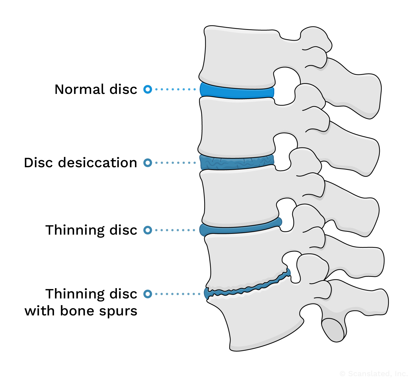
This diagram shows examples of degenerative changes in the spinal discs. With age or wear and tear, the spinal discs may start to dry out (dessication). This can sometimes progress to thinning of the disc and formation of bone spurs over time.
degenerative disc disease
डिजनरेटिव डिस्क बीमारी
उम्र के साथ रीढ़ की हड्डी की डिस्क का पतला होना और समय के साथ टूट-फूट होना।
स्पाइनल डिस्क रीढ़ की हड्डियों के बीच नरम, आघात-अवशोषित कुशन होते हैं जो रीढ़ को लचीला होने देते हैं।
डिस्क में टूट-फूट होना उम्र बढ़ने का एक सामान्य हिस्सा है, लेकिन परिवर्तन की मात्रा हर व्यक्ति में अलग-अलग होती है। चोट लगने या अधिक उपयोग के कारण युवा रोगियों में भी यह समस्या देखी जा सकती है।
डिस्क बीमारी (जिसे “डिस्क डीजनरेशन” भी कहा जाता है) पीठ दर्द का स्रोत हो भी सकता है और नहीं भी।
This diagram shows examples of degenerative changes in the spinal discs. With age or wear and tear, the spinal discs may start to dry out (dessication). This can sometimes progress to thinning of the disc and formation of bone spurs over time.
degenerative disc disease
discopatía degenerativa
Cambios en la columna vertebral asociados al envejecimiento y al desgaste que se produce a lo largo de los años; lo cual puede provocar el adelgazamiento de los discos que se encuentran entre los huesos de la columna vertebral. Esta situación es una parte normal del envejecimiento, pero también se puede observar en pacientes más jóvenes tras una lesión o por el uso excesivo. Los cambios varían de persona a persona, y puede que sean una causa del dolor de espalda.
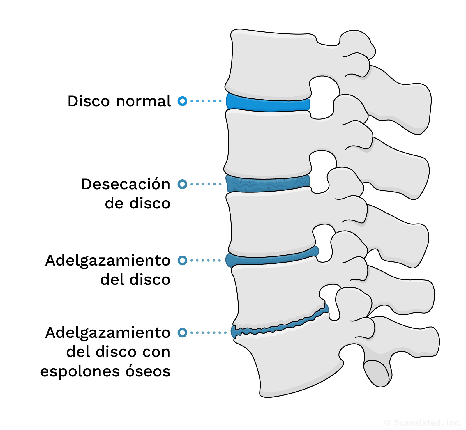
This diagram shows examples of degenerative changes in the spinal discs. With age or wear and tear, the spinal discs may start to dry out (dessication). This can sometimes progress to thinning of the disc and formation of bone spurs over time.
degenerative disc disease
椎间盘退行性疾病
由于随着时间推移逐渐磨损, 椎间盘发生与年龄相关的变薄和磨损。
椎间盘是椎骨之间的减震软垫, 使脊柱能够弯曲。
椎间盘的磨损性改变是衰老的正常现象, 但改变的程度因人而异。年轻患者也可能因为受伤或过度使用而出现这种情况。
椎间盘疾病(也称为”椎间盘退行性变”)可能是也可能不是背痛的根源。
This diagram shows examples of degenerative changes in the spinal discs. With age or wear and tear, the spinal discs may start to dry out (dessication). This can sometimes progress to thinning of the disc and formation of bone spurs over time.
degenerative disc disease
discopathie dégénérative
Amincissement et usure des disques vertébraux liés à l’âge, en raison de l’usure du temps.
Les disques vertébraux sont des coussins souples qui absorbent les chocs entre les os de la colonne vertébrale et qui permettent à cette dernière de se courber.
Les modifications dues à l’usure des disques sont un processus normal de vieillissement, mais l’ampleur de ces changements varie d’une personne à l’autre. Ce phénomène peut également être observé chez des patients plus jeunes à la suite d’une blessure ou d’une surutilisation.
La discopathie (également appelée « dégénérescence discale ») peut ou non être à l’origine de douleurs dorsales.
This diagram shows examples of degenerative changes in the spinal discs. With age or wear and tear, the spinal discs may start to dry out (dessication). This can sometimes progress to thinning of the disc and formation of bone spurs over time.
facet arthropathy
Arthritis or wear-and-tear changes of the small joints (facets) that link the spine bones (vertebrae) together and allow the spine to bend forward and backward.
These changes are common with age, and can lead to the narrowing of spaces around spinal nerves.
This is a general diagram showing facet joints in their normal state. It shows the lumbar section of the spine as an example.
facet arthropathy
फेसेट आर्थ्रोपैथी
गठिया या छोटे जोड़ों (पहलू) में होने वाले घिसावट और परिवर्तन, जो रीढ़ की हड्डियों (कशेरुक) को एक साथ जोड़ते हैं और रीढ़ को आगे और पीछे झुकने की अनुमति देते हैं।
ये परिवर्तन उम्र के साथ आम हैं, और इससे रीढ़ की हड्डी की नसों के आसपास की जगह सिकुड़ सकती है।
This is a general diagram showing facet joints in their normal state. It shows the lumbar section of the spine as an example.
facet arthropathy
artropatía facetaria
Artritis o cambios de desgaste de las articulaciones pequeñas que unen los huesos de la columna vertebral (vertebras). Normalmente, esto significa una reducción del espacio en las articulaciones (por el adelgazamiento del cartílago) o por la formación de espolones óseos a lo largo de la articulación. Estos cambios son habituales con el envejecimiento.
This is a general diagram showing facet joints in their normal state. It shows the lumbar section of the spine as an example.
facet arthropathy
关节面病变
连接脊椎骨(椎骨), 使得脊柱可以前后弯曲的小关节(关节面)发生关节炎或磨损性病变。
随着年龄的增长, 这些变化很常见, 并可能导致脊神经周围的空间变窄。
This is a general diagram showing facet joints in their normal state. It shows the lumbar section of the spine as an example.
facet arthropathy
arthropathie des facettes
Arthrite ou usure des petites articulations (facettes) qui relient les os de la colonne vertébrale (vertèbres) entre eux et permettent à la colonne vertébrale de se plier en avant et en arrière.
Ces changements sont fréquents avec l’âge et peuvent entraîner un rétrécissement des espaces autour des nerfs rachidiens.
This is a general diagram showing facet joints in their normal state. It shows the lumbar section of the spine as an example.
most pronounced
Most obvious or noticeable
most pronounced
सर्वाधिक स्पष्ट
सबसे स्पष्ट या ध्यान देने योग्य
most pronounced
más pronunciado
más notable
most pronounced
最明显的
最明显或最引人注目
most pronounced
le plus prononcé
Le plus évident ou le plus perceptible
L4-L5
At the level of the 4th and 5th lumbar vertebrae (spine bones) in the lower back.
There are usually five bones in the lumbar spine, which begins just below the rib cage and continues down through the lower back.
L4-L5
L4-L5
निचली पीठ में चौथी और पांचवीं काठ कशेरुका (रीढ़ की हड्डी) के स्तर पर।
काठ की रीढ़ में आमतौर पर पांच हड्डियां होती हैं, जो पसलियों के पिंजरे के ठीक नीचे से शुरू होती हैं और पीठ के निचले हिस्से से नीचे तक जाती हैं।
L4-L5
L4-L5
Entre los cuerpos vertebrales lumbares 4º y 5º (huesos de la parte baja de la espalda). Hay un disco (cojín amortiguador blando) entre los dos huesos y las articulaciones a lo largo de la parte posterior que une los huesos.
L4-L5
L4-L5
在下腰部第4和第5腰椎(脊椎骨)的位置。
腰椎通常有五块骨头, 从肋骨下方开始, 一直延伸到下背部。
L4-L5
L4-L5
Au niveau des 4ème et 5ème vertèbres lombaires (os de la colonne vertébrale) dans le bas du dos.
La colonne vertébrale lombaire, qui commence juste en dessous de la cage thoracique et se poursuit jusqu’au bas du dos, compte généralement cinq os.
L5-S1
Where the last lumbar vertebra (L5) meets the top of the sacrum (S1).
The sacrum is the large, triangle-shaped “sitting bone” that makes up the back part of the pelvis and is connected to the lower (lumbar) spine. It’s made up of five bones, which usually fuse (join together) during early adulthood.
L5-S1
L5-S1
जहां अंतिम कटि कशेरुका (L5) त्रिकास्थि (S1) के शीर्ष से मिलती है।
त्रिकास्थि बड़ी, त्रिकोण के आकार की “बैठने वाली हड्डी” है जो श्रोणि के पीछे का हिस्सा बनाती है और निचली (काठ) रीढ़ से जुड़ी होती है। यह पाँच हड्डियों से बनी होती है, जो आमतौर पर प्रारंभिक वयस्कता के दौरान जुड़ जाती हैं (एक साथ जुड़ जाती हैं)।
L5-S1
L5-S1
El espacio entre el último hueso lumbar (L5) y el primer hueso sacro (S1). Hay un disco (cojín amortiguador blando) entre los dos huesos y las articulaciones a lo largo de la parte posterior que une los huesos. El nivel L5-S1 se encuentra en la parte más baja de la espalda, justo por encima de los huesos del asiento.
L5-S1
L5-S1
最后一节腰椎(L5)与骶骨顶部(S1)的交接处。
骶骨是一块三角形的大”坐骨”, 构成骨盆的后部, 与下部(腰椎)脊柱相连。它由五块骨头组成, 通常在成年早期会融合(连接)在一起。
L5-S1
L5-S1
Point de rencontre entre la dernière vertèbre lombaire (L5) et la partie supérieure du sacrum (S1).
Le sacrum est le grand « os de l’assise » en forme de triangle qui constitue la partie arrière du bassin et est relié à la colonne vertébrale inférieure (lombaire). Il est composé de cinq os, qui fusionnent généralement au début de l’âge adulte.
bilateral
Involving or referring to both the left and right sides
neural foraminal narrowing
Narrowing (stenosis) of the neural foramina, which can be caused by bone spurs or bulging discs.
A “foramen” (plural: “foramina”) is a normal hole or tunnel in a bone.
In the spine, neural foramina are openings on either side of the vertebrae (spine bones) where the nerves travel. Nerves exit the spinal canal through these openings at each level of the spine, going out to the left and right sides of the body.
Narrowing of these openings can sometimes be a cause for pain.
neural foraminal narrowing
तंत्रिका छिद्र का संकुचन
तंत्रिका छिद्रों का संकुचित होना (स्टेनोसिस), जो अस्थि स्पर्स या उभरी हुई डिस्क के कारण हो सकता है।
एक “फोरैमन” (बहुवचन: “फोरैमिना”) हड्डी में एक सामान्य छेद या सुरंग है।
रीढ़ में, न्यूरल फोरैमिना कशेरुकाओं (रीढ़ की हड्डियों) के दोनों ओर खुले होते हैं जहां तंत्रिकाएं यात्रा करती हैं। रीढ़ की हड्डी के प्रत्येक स्तर पर इन छिद्रों के माध्यम से नसें रीढ़ की हड्डी की नहर से बाहर निकलती हैं, शरीर के बाएँ और दाएँ तरफ जाती हैं।
इन छिद्रों का संकुचित होना कभी-कभी दर्द का कारण बन सकता है।
neural foraminal narrowing
pinzamiento del agujero intervertebral
Estrechez de las aberturas en cada lado de los huesos de la columna vertebral por donde recorren los nervios. Los nervios salen del canal espinal por cada nivel de la columna vertebral y transmiten señales de movimiento y de sensación a las diferentes partes del cuerpo. A cada nivel, hay un nervio que se dirige al lado derecho, y un nervio que se dirige al lado izquierdo del cuerpo. Estos nervios recorren por el agujero intervertebral en cada lado al salir de la columna vertebral. El estrechamiento de estas aberturas puede ser causado por espolones óseos o hernias discales.
neural foraminal narrowing
神经孔狭窄
神经孔变窄(狭窄), 可能由骨刺或椎间盘膨出引起。
骨骼中存在正常的孔洞或隧道, 称之为”孔”。
在脊柱中, 神经孔是脊柱骨(椎骨)一侧的有神经穿过的开口。神经通过每个脊柱水平的这些开口离开椎管, 通向身体的左右两侧。
这些开口变窄有时会导致疼痛。
neural foraminal narrowing
rétrécissement du foramen neural
Le rétrécissement (sténose) du foramen neural peut être causé par des épines osseuses ou des hernies discales.
Le « foramen » (pluriel : « foramina » ) est un trou ou un tunnel normal dans un os.
Dans la colonne vertébrale, les foramina neuraux sont des ouvertures de chaque côté des vertèbres (les os de la colonne vertébrale) où passent les nerfs. Les nerfs sortent du canal rachidien par ces ouvertures à chaque niveau de la colonne vertébrale et partent à droite et à gauche du corps.
Le rétrécissement de ces ouvertures peut parfois provoquer de la douleur.
L3-L4
At the level of the 3rd and 4th lumbar vertebrae (spine bones) in the lower back.
There are usually five bones in the lumbar spine, which begins just below the rib cage and continues down through the lower back.
L3-L4
L3-L4
पीठ के निचले हिस्से में तीसरी और चौथी काठ कशेरुका (रीढ़ की हड्डियाँ) के स्तर पर।
काठ की रीढ़ में आमतौर पर पांच हड्डियां होती हैं, जो पसलियों के पिंजरे के ठीक नीचे से शुरू होती हैं और पीठ के निचले हिस्से से नीचे तक जाती हैं।
L3-L4
L3-L4
Entre la 3ª y 4ª vértebra lumbar (huesos de la parte baja de la espalda). Hay un disco (cojín amortiguador blando) entre los dos huesos y las articulaciones a lo largo de la parte posterior que une los huesos.
L3-L4
L3-L4
位于下腰部第3和第4腰椎(脊椎骨)的位置。
腰椎通常有五块骨头, 从肋骨下方开始, 一直延伸到下背部。
L3-L4
L3-L4
Au niveau des 3ème et 4ème vertèbres lombaires (os de la colonne vertébrale) dans le bas du dos.
La colonne vertébrale lombaire, qui commence juste en dessous de la cage thoracique et se poursuit jusqu’au bas du dos, compte généralement cinq os.
L5-S1
Where the last lumbar vertebra (L5) meets the top of the sacrum (S1).
The sacrum is the large, triangle-shaped “sitting bone” that makes up the back part of the pelvis and is connected to the lower (lumbar) spine. It’s made up of five bones, which usually fuse (join together) during early adulthood.
L5-S1
L5-S1
जहां अंतिम कटि कशेरुका (L5) त्रिकास्थि (S1) के शीर्ष से मिलती है।
त्रिकास्थि बड़ी, त्रिकोण के आकार की “बैठने वाली हड्डी” है जो श्रोणि के पीछे का हिस्सा बनाती है और निचली (काठ) रीढ़ से जुड़ी होती है। यह पाँच हड्डियों से बनी होती है, जो आमतौर पर प्रारंभिक वयस्कता के दौरान जुड़ जाती हैं (एक साथ जुड़ जाती हैं)।
L5-S1
L5-S1
El espacio entre el último hueso lumbar (L5) y el primer hueso sacro (S1). Hay un disco (cojín amortiguador blando) entre los dos huesos y las articulaciones a lo largo de la parte posterior que une los huesos. El nivel L5-S1 se encuentra en la parte más baja de la espalda, justo por encima de los huesos del asiento.
L5-S1
L5-S1
最后一节腰椎(L5)与骶骨顶部(S1)的交接处。
骶骨是一块三角形的大”坐骨”, 构成骨盆的后部, 与下部(腰椎)脊柱相连。它由五块骨头组成, 通常在成年早期会融合(连接)在一起。
L5-S1
L5-S1
Point de rencontre entre la dernière vertèbre lombaire (L5) et la partie supérieure du sacrum (S1).
Le sacrum est le grand « os de l’assise » en forme de triangle qui constitue la partie arrière du bassin et est relié à la colonne vertébrale inférieure (lombaire). Il est composé de cinq os, qui fusionnent généralement au début de l’âge adulte.
Radiologist
A doctor trained in the field of medical imaging, to supervise and interpret radiology exams
